+ Open data
- Basic information
| Entry | Database: PDB / ID: 8fi0 | ||||||
|---|---|---|---|---|---|---|---|
| Title | Structure of Lettuce aptamer bound to DFAME | ||||||
 Components Components | Lettuce DNA aptamer | ||||||
 Keywords Keywords | DNA / fluorescence / turn-on / fluorogenic / fluorophore / G-quartet / G-quadruplex | ||||||
| Function / homology | : / Chem-X5R / DNA / DNA (> 10) Function and homology information Function and homology information | ||||||
| Biological species | synthetic construct (others) | ||||||
| Method |  X-RAY DIFFRACTION / X-RAY DIFFRACTION /  SYNCHROTRON / SYNCHROTRON /  MOLECULAR REPLACEMENT / Resolution: 3 Å MOLECULAR REPLACEMENT / Resolution: 3 Å | ||||||
 Authors Authors | Passalacqua, L.F.M. / Ferre-D'Amare, A.R. | ||||||
| Funding support |  United States, 1items United States, 1items
| ||||||
 Citation Citation |  Journal: Nature / Year: 2023 Journal: Nature / Year: 2023Title: Intricate 3D architecture of a DNA mimic of GFP. Authors: Luiz F M Passalacqua / Michael T Banco / Jared D Moon / Xing Li / Samie R Jaffrey / Adrian R Ferré-D'Amaré /   Abstract: Numerous studies have shown how RNA molecules can adopt elaborate three-dimensional (3D) architectures. By contrast, whether DNA can self-assemble into complex 3D folds capable of sophisticated ...Numerous studies have shown how RNA molecules can adopt elaborate three-dimensional (3D) architectures. By contrast, whether DNA can self-assemble into complex 3D folds capable of sophisticated biochemistry, independent of protein or RNA partners, has remained mysterious. Lettuce is an in vitro-evolved DNA molecule that binds and activates conditional fluorophores derived from GFP. To extend previous structural studies of fluorogenic RNAs, GFP and other fluorescent proteins to DNA, we characterize Lettuce-fluorophore complexes by X-ray crystallography and cryogenic electron microscopy. The results reveal that the 53-nucleotide DNA adopts a four-way junction (4WJ) fold. Instead of the canonical L-shaped or H-shaped structures commonly seen in 4WJ RNAs, the four stems of Lettuce form two coaxial stacks that pack co-linearly to form a central G-quadruplex in which the fluorophore binds. This fold is stabilized by stacking, extensive nucleobase hydrogen bonding-including through unusual diagonally stacked bases that bridge successive tiers of the main coaxial stacks of the DNA-and coordination of monovalent and divalent cations. Overall, the structure is more compact than many RNAs of comparable size. Lettuce demonstrates how DNA can form elaborate 3D structures without using RNA-like tertiary interactions and suggests that new principles of nucleic acid organization will be forthcoming from the analysis of complex DNAs. | ||||||
| History |
|
- Structure visualization
| Structure viewer | Molecule:  Molmil Molmil Jmol/JSmol Jmol/JSmol |
|---|
- Downloads & links
- Download
| PDBx/mmCIF format |  8fi0.cif.gz 8fi0.cif.gz | 85.4 KB | Display |  PDBx/mmCIF format PDBx/mmCIF format |
|---|---|---|---|---|
| PDB format |  pdb8fi0.ent.gz pdb8fi0.ent.gz | 53.6 KB | Display |  PDB format PDB format |
| PDBx/mmJSON format |  8fi0.json.gz 8fi0.json.gz | Tree view |  PDBx/mmJSON format PDBx/mmJSON format | |
| Others |  Other downloads Other downloads |
-Validation report
| Summary document |  8fi0_validation.pdf.gz 8fi0_validation.pdf.gz | 629.7 KB | Display |  wwPDB validaton report wwPDB validaton report |
|---|---|---|---|---|
| Full document |  8fi0_full_validation.pdf.gz 8fi0_full_validation.pdf.gz | 632.7 KB | Display | |
| Data in XML |  8fi0_validation.xml.gz 8fi0_validation.xml.gz | 4.6 KB | Display | |
| Data in CIF |  8fi0_validation.cif.gz 8fi0_validation.cif.gz | 5.7 KB | Display | |
| Arichive directory |  https://data.pdbj.org/pub/pdb/validation_reports/fi/8fi0 https://data.pdbj.org/pub/pdb/validation_reports/fi/8fi0 ftp://data.pdbj.org/pub/pdb/validation_reports/fi/8fi0 ftp://data.pdbj.org/pub/pdb/validation_reports/fi/8fi0 | HTTPS FTP |
-Related structure data
| Related structure data |  8fhvSC 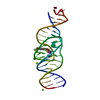 8fhxC 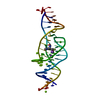 8fhzC 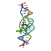 8fi1C 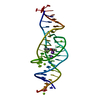 8fi2C  8fi7C  8fi8C S: Starting model for refinement C: citing same article ( |
|---|---|
| Similar structure data | Similarity search - Function & homology  F&H Search F&H Search |
- Links
Links
- Assembly
| Deposited unit | 
| ||||||||||||
|---|---|---|---|---|---|---|---|---|---|---|---|---|---|
| 1 |
| ||||||||||||
| Unit cell |
|
- Components
| #1: DNA chain | Mass: 16542.562 Da / Num. of mol.: 1 / Source method: obtained synthetically / Source: (synth.) synthetic construct (others) | ||||||||
|---|---|---|---|---|---|---|---|---|---|
| #2: Chemical | | #3: Chemical | ChemComp-MG / #4: Chemical | ChemComp-X5R / | #5: Water | ChemComp-HOH / | Has ligand of interest | Y | |
-Experimental details
-Experiment
| Experiment | Method:  X-RAY DIFFRACTION / Number of used crystals: 1 X-RAY DIFFRACTION / Number of used crystals: 1 |
|---|
- Sample preparation
| Crystal | Density Matthews: 1.93 Å3/Da / Density % sol: 36.43 % |
|---|---|
| Crystal grow | Temperature: 294 K / Method: vapor diffusion, sitting drop / pH: 7.5 Details: 0.05 M Magnesium chloride hexahydrate, 0.1 M HEPES pH 7.5, 30% (v/v) polyethylene glycol monoethyl ether 550 |
-Data collection
| Diffraction | Mean temperature: 100 K / Serial crystal experiment: N |
|---|---|
| Diffraction source | Source:  SYNCHROTRON / Site: SYNCHROTRON / Site:  ALS ALS  / Beamline: 5.0.1 / Wavelength: 0.977 Å / Beamline: 5.0.1 / Wavelength: 0.977 Å |
| Detector | Type: DECTRIS PILATUS3 2M / Detector: PIXEL / Date: Aug 30, 2022 |
| Radiation | Protocol: SINGLE WAVELENGTH / Monochromatic (M) / Laue (L): M / Scattering type: x-ray |
| Radiation wavelength | Wavelength: 0.977 Å / Relative weight: 1 |
| Reflection | Resolution: 2.9→59.81 Å / Num. obs: 3164 / % possible obs: 99.91 % / Redundancy: 11.46 % / Biso Wilson estimate: 56.84 Å2 / CC1/2: 0.994 / Rmerge(I) obs: 0.07 / Net I/σ(I): 20 |
| Reflection shell | Resolution: 2.9→3 Å / Rmerge(I) obs: 0.269 / Mean I/σ(I) obs: 3.1 / Num. unique obs: 300 / CC1/2: 0.987 |
- Processing
| Software |
| ||||||||||||||||||||||||||||||||||||||||
|---|---|---|---|---|---|---|---|---|---|---|---|---|---|---|---|---|---|---|---|---|---|---|---|---|---|---|---|---|---|---|---|---|---|---|---|---|---|---|---|---|---|
| Refinement | Method to determine structure:  MOLECULAR REPLACEMENT MOLECULAR REPLACEMENTStarting model: 8FHV Resolution: 3→59.81 Å / SU ML: 0.4108 / Cross valid method: FREE R-VALUE / σ(F): 1.44 / Phase error: 19.6322 Stereochemistry target values: GeoStd + Monomer Library + CDL v1.2
| ||||||||||||||||||||||||||||||||||||||||
| Solvent computation | Shrinkage radii: 0.9 Å / VDW probe radii: 1.11 Å / Solvent model: FLAT BULK SOLVENT MODEL | ||||||||||||||||||||||||||||||||||||||||
| Displacement parameters | Biso mean: 52.68 Å2 | ||||||||||||||||||||||||||||||||||||||||
| Refinement step | Cycle: LAST / Resolution: 3→59.81 Å
| ||||||||||||||||||||||||||||||||||||||||
| Refine LS restraints |
| ||||||||||||||||||||||||||||||||||||||||
| LS refinement shell |
| ||||||||||||||||||||||||||||||||||||||||
| Refinement TLS params. | Method: refined / Origin x: 6.10049284811 Å / Origin y: -39.6878051661 Å / Origin z: -102.799266861 Å
| ||||||||||||||||||||||||||||||||||||||||
| Refinement TLS group | Selection details: all |
 Movie
Movie Controller


 PDBj
PDBj


