+ Open data
- Basic information
| Entry |  | |||||||||
|---|---|---|---|---|---|---|---|---|---|---|
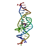
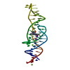
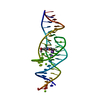
| Title | Cryo-EM map of the unliganded Lettuce aptamer | |||||||||
 Map data Map data | Map of file (mrc format) for apo Lettuce | |||||||||
 Sample Sample |
| |||||||||
 Keywords Keywords | DNA / aptamer / fluorescence / turn-on / fluorogenic / fluorophore | |||||||||
| Method | single particle reconstruction / cryo EM / Resolution: 6.5 Å | |||||||||
 Authors Authors | Banco MT / Passalacqua LFM / Ferre-D'Amare AR | |||||||||
| Funding support |  United States, 1 items United States, 1 items
| |||||||||
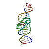
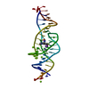
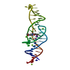
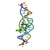
 Citation Citation |  Journal: Nature / Year: 2023 Journal: Nature / Year: 2023Title: Intricate 3D architecture of a DNA mimic of GFP. Authors: Luiz F M Passalacqua / Michael T Banco / Jared D Moon / Xing Li / Samie R Jaffrey / Adrian R Ferré-D'Amaré /   Abstract: Numerous studies have shown how RNA molecules can adopt elaborate three-dimensional (3D) architectures. By contrast, whether DNA can self-assemble into complex 3D folds capable of sophisticated ...Numerous studies have shown how RNA molecules can adopt elaborate three-dimensional (3D) architectures. By contrast, whether DNA can self-assemble into complex 3D folds capable of sophisticated biochemistry, independent of protein or RNA partners, has remained mysterious. Lettuce is an in vitro-evolved DNA molecule that binds and activates conditional fluorophores derived from GFP. To extend previous structural studies of fluorogenic RNAs, GFP and other fluorescent proteins to DNA, we characterize Lettuce-fluorophore complexes by X-ray crystallography and cryogenic electron microscopy. The results reveal that the 53-nucleotide DNA adopts a four-way junction (4WJ) fold. Instead of the canonical L-shaped or H-shaped structures commonly seen in 4WJ RNAs, the four stems of Lettuce form two coaxial stacks that pack co-linearly to form a central G-quadruplex in which the fluorophore binds. This fold is stabilized by stacking, extensive nucleobase hydrogen bonding-including through unusual diagonally stacked bases that bridge successive tiers of the main coaxial stacks of the DNA-and coordination of monovalent and divalent cations. Overall, the structure is more compact than many RNAs of comparable size. Lettuce demonstrates how DNA can form elaborate 3D structures without using RNA-like tertiary interactions and suggests that new principles of nucleic acid organization will be forthcoming from the analysis of complex DNAs. | |||||||||
| History |
|
- Structure visualization
| Supplemental images |
|---|
- Downloads & links
-EMDB archive
| Map data |  emd_29329.map.gz emd_29329.map.gz | 25.6 MB |  EMDB map data format EMDB map data format | |
|---|---|---|---|---|
| Header (meta data) |  emd-29329-v30.xml emd-29329-v30.xml emd-29329.xml emd-29329.xml | 13.3 KB 13.3 KB | Display Display |  EMDB header EMDB header |
| FSC (resolution estimation) |  emd_29329_fsc.xml emd_29329_fsc.xml | 9 KB | Display |  FSC data file FSC data file |
| Images |  emd_29329.png emd_29329.png | 22.6 KB | ||
| Others |  emd_29329_half_map_1.map.gz emd_29329_half_map_1.map.gz emd_29329_half_map_2.map.gz emd_29329_half_map_2.map.gz | 48.9 MB 48.9 MB | ||
| Archive directory |  http://ftp.pdbj.org/pub/emdb/structures/EMD-29329 http://ftp.pdbj.org/pub/emdb/structures/EMD-29329 ftp://ftp.pdbj.org/pub/emdb/structures/EMD-29329 ftp://ftp.pdbj.org/pub/emdb/structures/EMD-29329 | HTTPS FTP |
-Validation report
| Summary document |  emd_29329_validation.pdf.gz emd_29329_validation.pdf.gz | 635.5 KB | Display |  EMDB validaton report EMDB validaton report |
|---|---|---|---|---|
| Full document |  emd_29329_full_validation.pdf.gz emd_29329_full_validation.pdf.gz | 635.1 KB | Display | |
| Data in XML |  emd_29329_validation.xml.gz emd_29329_validation.xml.gz | 15.9 KB | Display | |
| Data in CIF |  emd_29329_validation.cif.gz emd_29329_validation.cif.gz | 20.4 KB | Display | |
| Arichive directory |  https://ftp.pdbj.org/pub/emdb/validation_reports/EMD-29329 https://ftp.pdbj.org/pub/emdb/validation_reports/EMD-29329 ftp://ftp.pdbj.org/pub/emdb/validation_reports/EMD-29329 ftp://ftp.pdbj.org/pub/emdb/validation_reports/EMD-29329 | HTTPS FTP |
-Related structure data
- Links
Links
| EMDB pages |  EMDB (EBI/PDBe) / EMDB (EBI/PDBe) /  EMDataResource EMDataResource |
|---|
- Map
Map
| File |  Download / File: emd_29329.map.gz / Format: CCP4 / Size: 52.7 MB / Type: IMAGE STORED AS FLOATING POINT NUMBER (4 BYTES) Download / File: emd_29329.map.gz / Format: CCP4 / Size: 52.7 MB / Type: IMAGE STORED AS FLOATING POINT NUMBER (4 BYTES) | ||||||||||||||||||||||||||||||||||||
|---|---|---|---|---|---|---|---|---|---|---|---|---|---|---|---|---|---|---|---|---|---|---|---|---|---|---|---|---|---|---|---|---|---|---|---|---|---|
| Annotation | Map of file (mrc format) for apo Lettuce | ||||||||||||||||||||||||||||||||||||
| Projections & slices | Image control
Images are generated by Spider. | ||||||||||||||||||||||||||||||||||||
| Voxel size | X=Y=Z: 0.9 Å | ||||||||||||||||||||||||||||||||||||
| Density |
| ||||||||||||||||||||||||||||||||||||
| Symmetry | Space group: 1 | ||||||||||||||||||||||||||||||||||||
| Details | EMDB XML:
|
-Supplemental data
-Half map: Half map A
| File | emd_29329_half_map_1.map | ||||||||||||
|---|---|---|---|---|---|---|---|---|---|---|---|---|---|
| Annotation | Half map A | ||||||||||||
| Projections & Slices |
| ||||||||||||
| Density Histograms |
-Half map: Half map B
| File | emd_29329_half_map_2.map | ||||||||||||
|---|---|---|---|---|---|---|---|---|---|---|---|---|---|
| Annotation | Half map B | ||||||||||||
| Projections & Slices |
| ||||||||||||
| Density Histograms |
- Sample components
-Entire : Lettuce
| Entire | Name: Lettuce |
|---|---|
| Components |
|
-Supramolecule #1: Lettuce
| Supramolecule | Name: Lettuce / type: complex / ID: 1 / Parent: 0 / Details: Synthetic in vitro evolved DNA aptamer |
|---|
-Experimental details
-Structure determination
| Method | cryo EM |
|---|---|
 Processing Processing | single particle reconstruction |
| Aggregation state | particle |
- Sample preparation
| Buffer | pH: 7 |
|---|---|
| Grid | Model: Quantifoil R1.2/1.3 / Material: COPPER / Mesh: 300 / Support film - Material: CARBON / Support film - topology: HOLEY / Pretreatment - Type: GLOW DISCHARGE |
| Vitrification | Cryogen name: ETHANE / Chamber humidity: 100 % / Instrument: FEI VITROBOT MARK IV |
- Electron microscopy
| Microscope | TFS GLACIOS |
|---|---|
| Image recording | Film or detector model: FEI FALCON IV (4k x 4k) / Number real images: 3327 / Average electron dose: 52.5 e/Å2 |
| Electron beam | Acceleration voltage: 200 kV / Electron source:  FIELD EMISSION GUN FIELD EMISSION GUN |
| Electron optics | Illumination mode: SPOT SCAN / Imaging mode: BRIGHT FIELD / Nominal defocus max: 2.2 µm / Nominal defocus min: 0.8 µm |
 Movie
Movie Controller


 Z (Sec.)
 Y (Row.)
 X (Col.)


