[English] 日本語
Yorodumi
- PDB-8fi7: Structure of Lettuce C20T bound to DFHO -

+
Open data


ID or keywords:

Loading...

-
Basic information

Entry
Database: PDB / ID: 8fi7
TitleStructure of Lettuce C20T bound to DFHO
ComponentsLettuce DNA aptamer
KeywordsDNA / aptamer / fluorescence / turn-on / fluorogenic / fluorophore / G-quartet / G-quadruplex
Function / homologyChem-747 / : / DNA / DNA (> 10)
Function and homology information
Biological speciessynthetic construct (others)
MethodX-RAY DIFFRACTION / SYNCHROTRON / MOLECULAR REPLACEMENT / Resolution: 2.9 Å
AuthorsPassalacqua, L.F.M. / Ferre-D'Amare, A.R.
Funding support United States, 1items
OrganizationGrant numberCountry
National Institutes of Health/National Heart, Lung, and Blood Institute (NIH/NHLBI) United States
Citation
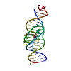
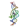
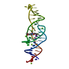
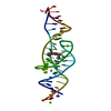
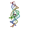
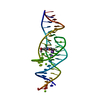
Journal: Nature / Year: 2023
Title: Intricate 3D architecture of a DNA mimic of GFP.
Authors: Luiz F M Passalacqua / Michael T Banco / Jared D Moon / Xing Li / Samie R Jaffrey / Adrian R Ferré-D'Amaré /
Abstract: Numerous studies have shown how RNA molecules can adopt elaborate three-dimensional (3D) architectures. By contrast, whether DNA can self-assemble into complex 3D folds capable of sophisticated ...Numerous studies have shown how RNA molecules can adopt elaborate three-dimensional (3D) architectures. By contrast, whether DNA can self-assemble into complex 3D folds capable of sophisticated biochemistry, independent of protein or RNA partners, has remained mysterious. Lettuce is an in vitro-evolved DNA molecule that binds and activates conditional fluorophores derived from GFP. To extend previous structural studies of fluorogenic RNAs, GFP and other fluorescent proteins to DNA, we characterize Lettuce-fluorophore complexes by X-ray crystallography and cryogenic electron microscopy. The results reveal that the 53-nucleotide DNA adopts a four-way junction (4WJ) fold. Instead of the canonical L-shaped or H-shaped structures commonly seen in 4WJ RNAs, the four stems of Lettuce form two coaxial stacks that pack co-linearly to form a central G-quadruplex in which the fluorophore binds. This fold is stabilized by stacking, extensive nucleobase hydrogen bonding-including through unusual diagonally stacked bases that bridge successive tiers of the main coaxial stacks of the DNA-and coordination of monovalent and divalent cations. Overall, the structure is more compact than many RNAs of comparable size. Lettuce demonstrates how DNA can form elaborate 3D structures without using RNA-like tertiary interactions and suggests that new principles of nucleic acid organization will be forthcoming from the analysis of complex DNAs.
History
DepositionDec 15, 2022Deposition site: RCSB / Processing site: RCSB
Revision 1.0May 10, 2023Provider: repository / Type: Initial release
Revision 1.1Jul 5, 2023Group: Database references / Category: citation / citation_author
Item: _citation.country / _citation.journal_abbrev ..._citation.country / _citation.journal_abbrev / _citation.journal_id_ASTM / _citation.journal_id_CSD / _citation.journal_id_ISSN / _citation.journal_volume / _citation.page_first / _citation.page_last / _citation.pdbx_database_id_DOI / _citation.pdbx_database_id_PubMed / _citation.title / _citation.year
Revision 1.2Oct 25, 2023Group: Data collection / Category: chem_comp_atom / chem_comp_bond

-
Structure visualization

Structure viewerMolecule:
MolmilJmol/JSmol

Downloads & links

-
Assembly

Deposited unit
A: Lettuce DNA aptamer
hetero molecules


Theoretical massNumber of molelcules
Total (without water)16,9907
Polymers16,5581
Non-polymers4326
Water28816
1


  • Idetical with deposited unit
  • defined by author
  • Evidence: gel filtration
TypeNameSymmetry operationNumber
identity operation1_555x,y,z1
Unit cell
Length a, b, c (Å)24.571, 43.104, 117.641
Angle α, β, γ (deg.)90.000, 90.000, 90.000
Int Tables number19
Space group name H-MP212121
Space group name HallP2ac2ab
Symmetry operation#1: x,y,z
#2: x+1/2,-y+1/2,-z
#3: -x,y+1/2,-z+1/2
#4: -x+1/2,-y,z+1/2

-
Components

#1: DNA chain Lettuce DNA aptamer


Mass: 16557.574 Da / Num. of mol.: 1 / Mutation: C20T / Source method: obtained synthetically / Source: (synth.) synthetic construct (others)
#2: Chemical ChemComp-K / POTASSIUM ION


Mass: 39.098 Da / Num. of mol.: 2 / Source method: obtained synthetically / Formula: K
#3: Chemical ChemComp-MG / MAGNESIUM ION


Mass: 24.305 Da / Num. of mol.: 3 / Source method: obtained synthetically / Formula: Mg
#4: Chemical ChemComp-747 / (5Z)-5-[(3,5-difluoro-4-hydroxyphenyl)methylidene]-2-[(E)-(hydroxyimino)methyl]-3-methyl-3,5-dihydro-4H-imidazol-4-one


Mass: 281.215 Da / Num. of mol.: 1 / Source method: obtained synthetically / Formula: C12H9F2N3O3 / Feature type: SUBJECT OF INVESTIGATION
#5: Water ChemComp-HOH / water / Water


Mass: 18.015 Da / Num. of mol.: 16 / Source method: isolated from a natural source / Formula: H2O
Has ligand of interestY

-
Experimental details

-
Experiment

ExperimentMethod: X-RAY DIFFRACTION / Number of used crystals: 1

-
Sample preparation

CrystalDensity Matthews: 1.88 Å3/Da / Density % sol: 34.62 %
Crystal growTemperature: 294 K / Method: vapor diffusion, sitting drop / pH: 7.5
Details: 0.05 M Magnesium chloride hexahydrate, 0.1 M HEPES pH 7.5, 30% (v/v) polyethylene glycol monoethyl ether 550

-
Data collection

DiffractionMean temperature: 100 K / Serial crystal experiment: N
Diffraction sourceSource: SYNCHROTRON / Site: APS / Beamline: 24-ID-C / Wavelength: 1.105 Å
DetectorType: DECTRIS EIGER2 X 16M / Detector: PIXEL / Date: Jun 24, 2022
RadiationProtocol: SINGLE WAVELENGTH / Monochromatic (M) / Laue (L): M / Scattering type: x-ray
Radiation wavelengthWavelength: 1.105 Å / Relative weight: 1
ReflectionResolution: 2.8→40.47 Å / Num. obs: 3374 / % possible obs: 98.63 % / Redundancy: 9.15 % / Biso Wilson estimate: 52.33 Å2 / CC1/2: 0.996 / Rmerge(I) obs: 0.164 / Net I/σ(I): 22.8
Reflection shellResolution: 2.8→2.9 Å / Rmerge(I) obs: 0.442 / Mean I/σ(I) obs: 2.5 / Num. unique obs: 325 / CC1/2: 0.958

-
Processing

Software
NameVersionClassification
Coot0.9.5model building
PHENIX1.19.2_4158refinement
xia2data reduction
Aimlessdata scaling
PHASERphasing
RefinementMethod to determine structure: MOLECULAR REPLACEMENT
Starting model: 8FHV
Resolution: 2.9→34.77 Å / SU ML: 0.378 / Cross valid method: FREE R-VALUE / σ(F): 1.43 / Phase error: 19.9776
Stereochemistry target values: GeoStd + Monomer Library + CDL v1.2
RfactorNum. reflection% reflection
Rfree0.289 282 9.37 %
Rwork0.244 2727 -
obs0.2471 3048 97.73 %
Solvent computationShrinkage radii: 0.9 Å / VDW probe radii: 1.11 Å / Solvent model: FLAT BULK SOLVENT MODEL
Displacement parametersBiso mean: 44.99 Å2
Refinement stepCycle: LAST / Resolution: 2.9→34.77 Å
ProteinNucleic acidLigandSolventTotal
Num. atoms0 1100 25 16 1141
Refine LS restraints
Refine-IDTypeDev idealNumber
X-RAY DIFFRACTIONf_bond_d0.00461282
X-RAY DIFFRACTIONf_angle_d0.67871982
X-RAY DIFFRACTIONf_chiral_restr0.0313215
X-RAY DIFFRACTIONf_plane_restr0.004457
X-RAY DIFFRACTIONf_dihedral_angle_d35.3843542
LS refinement shell
Resolution (Å)Rfactor RfreeNum. reflection RfreeRfactor RworkNum. reflection RworkRefine-ID% reflection obs (%)
2.9-3.650.32221360.24311300X-RAY DIFFRACTION96.38
3.65-34.770.26951460.24281427X-RAY DIFFRACTION98.99
Refinement TLS params.Method: refined / Origin x: -18.5317092337 Å / Origin y: -39.8640434732 Å / Origin z: -101.061750959 Å
111213212223313233
T0.494142000184 Å2-0.0217403442403 Å2-0.0556863700079 Å2-0.492500914149 Å2-0.0361802481194 Å2--0.0949260087338 Å2
L0.565769990036 °2-0.00981226619443 °2-0.0692646029429 °2-0.586255106012 °20.167141944 °2--0.465151781698 °2
S0.075404006507 Å °0.213753645389 Å °-0.0995553476789 Å °-0.222209740828 Å °0.122102845755 Å °0.0842676770388 Å °0.0551658248986 Å °-0.0580464935586 Å °0.210381491552 Å °
Refinement TLS groupSelection details: all

+
About Yorodumi

-
News

-
Feb 9, 2022. New format data for meta-information of EMDB entries

New format data for meta-information of EMDB entries

  • Version 3 of the EMDB header file is now the official format.
  • The previous official version 1.9 will be removed from the archive.

Related info.:EMDB header

External links:wwPDB to switch to version 3 of the EMDB data model

-
Aug 12, 2020. Covid-19 info

Covid-19 info

URL: https://pdbj.org/emnavi/covid19.php

New page: Covid-19 featured information page in EM Navigator.

Related info.:Covid-19 info / Mar 5, 2020. Novel coronavirus structure data

+
Mar 5, 2020. Novel coronavirus structure data

Novel coronavirus structure data

Related info.:Yorodumi Speices / Aug 12, 2020. Covid-19 info

External links:COVID-19 featured content - PDBj / Molecule of the Month (242):Coronavirus Proteases

+
Jan 31, 2019. EMDB accession codes are about to change! (news from PDBe EMDB page)

EMDB accession codes are about to change! (news from PDBe EMDB page)

  • The allocation of 4 digits for EMDB accession codes will soon come to an end. Whilst these codes will remain in use, new EMDB accession codes will include an additional digit and will expand incrementally as the available range of codes is exhausted. The current 4-digit format prefixed with “EMD-” (i.e. EMD-XXXX) will advance to a 5-digit format (i.e. EMD-XXXXX), and so on. It is currently estimated that the 4-digit codes will be depleted around Spring 2019, at which point the 5-digit format will come into force.
  • The EM Navigator/Yorodumi systems omit the EMD- prefix.

Related info.:Q: What is EMD? / ID/Accession-code notation in Yorodumi/EM Navigator

External links:EMDB Accession Codes are Changing Soon! / Contact to PDBj

+
Jul 12, 2017. Major update of PDB

Major update of PDB

  • wwPDB released updated PDB data conforming to the new PDBx/mmCIF dictionary.
  • This is a major update changing the version number from 4 to 5, and with Remediation, in which all the entries are updated.
  • In this update, many items about electron microscopy experimental information are reorganized (e.g. em_software).
  • Now, EM Navigator and Yorodumi are based on the updated data.

External links:wwPDB Remediation / Enriched Model Files Conforming to OneDep Data Standards Now Available in the PDB FTP Archive

-
Yorodumi

Thousand views of thousand structures

  • Yorodumi is a browser for structure data from EMDB, PDB, SASBDB, etc.
  • This page is also the successor to EM Navigator detail page, and also detail information page/front-end page for Omokage search.
  • The word "yorodu" (or yorozu) is an old Japanese word meaning "ten thousand". "mi" (miru) is to see.

Related info.:EMDB / PDB / SASBDB / Comparison of 3 databanks / Yorodumi Search / Aug 31, 2016. New EM Navigator & Yorodumi / Yorodumi Papers / Jmol/JSmol / Function and homology information / Changes in new EM Navigator and Yorodumi

Read more