 Movie
Movie Controller

[English] 日本語
 Yorodumi
- PDB-8ewt: Bile salt hydrolase A from Lactobacillus gasseri bound to covalen... -
+ Open data

- Basic information
| Entry | Database: PDB / ID: 8ewt | ||||||
|---|---|---|---|---|---|---|---|
| Title | Bile salt hydrolase A from Lactobacillus gasseri bound to covalent probe | ||||||
 Components Components | Conjugated bile salt hydrolase | ||||||
 Keywords Keywords | HYDROLASE / bile salt hydrolase | ||||||
| Function / homology |  Function and homology information Function and homology informationchenodeoxycholoyltaurine hydrolase / choloylglycine hydrolase / Hydrolases; Acting on carbon-nitrogen bonds, other than peptide bonds; In linear amides / lipid metabolic process / hydrolase activity Similarity search - Function | ||||||
| Biological species |  Lactobacillus gasseri (bacteria) Lactobacillus gasseri (bacteria) | ||||||
| Method |  X-RAY DIFFRACTION / SYNCHROTRON / MOLECULAR REPLACEMENT / Resolution: 2.03 Å X-RAY DIFFRACTION / SYNCHROTRON / MOLECULAR REPLACEMENT / Resolution: 2.03 Å | ||||||
 Authors Authors | Walker, M.E. / Redinbo, M.R. | ||||||
| Funding support |  United States, 1items United States, 1items
| ||||||
 Citation Citation | Journal: To Be Published Title: Structural diversity of bile salt hydrolases reveals rationale for substrate selectivity Authors: Walker, M.E. / Redinbo, M.R. | ||||||
| History |
|
- Structure visualization
| Structure viewer | Molecule: MolmilJmol/JSmol |
|---|
- Downloads & links
-Download
| PDBx/mmCIF format | 8ewt.cif.gz | 171.7 KB | Display | PDBx/mmCIF format |
|---|---|---|---|---|
| PDB format | pdb8ewt.ent.gz | 108.5 KB | Display | PDB format |
| PDBx/mmJSON format | 8ewt.json.gz | Tree view | PDBx/mmJSON format | |
| Others |  Other downloads Other downloads |
-Validation report
| Summary document | 8ewt_validation.pdf.gz | 944.9 KB | Display | wwPDB validaton report |
|---|---|---|---|---|
| Full document | 8ewt_full_validation.pdf.gz | 945.7 KB | Display | |
| Data in XML | 8ewt_validation.xml.gz | 24.5 KB | Display | |
| Data in CIF | 8ewt_validation.cif.gz | 35 KB | Display | |
| Arichive directory | https://data.pdbj.org/pub/pdb/validation_reports/ew/8ewtftp://data.pdbj.org/pub/pdb/validation_reports/ew/8ewt | HTTPS FTP |
-Related structure data
| Related structure data |  8esgC  8esiC  8eslC  8eteC  8etfC  8etkC  8faoC 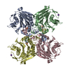 7svfS S: Starting model for refinement C: citing same article ( |
|---|---|
| Similar structure data |
-Links
 PDBj
PDBj


- Assembly
| Deposited unit | 
| ||||||||||||
|---|---|---|---|---|---|---|---|---|---|---|---|---|---|
| 1 | 
| ||||||||||||
| Unit cell |
|
-Components
| #1: Protein | Mass: 35860.262 Da / Num. of mol.: 2 Source method: isolated from a genetically manipulated source Source: (gene. exp.) Lactobacillus gasseri (bacteria) / Gene: LGAS_0054 / Production host: #2: Chemical |   Mass: 430.638 Da / Num. of mol.: 2 / Source method: obtained synthetically / Formula: C28H43FO2 / Feature type: SUBJECT OF INVESTIGATION Mass: 430.638 Da / Num. of mol.: 2 / Source method: obtained synthetically / Formula: C28H43FO2 / Feature type: SUBJECT OF INVESTIGATION#3: Chemical |   Mass: 22.990 Da / Num. of mol.: 2 / Source method: isolated from a natural source / Formula: Na Mass: 22.990 Da / Num. of mol.: 2 / Source method: isolated from a natural source / Formula: Na#4: Water | ChemComp-HOH / |  Mass: 18.015 Da / Num. of mol.: 178 / Source method: isolated from a natural source / Formula: H2O Mass: 18.015 Da / Num. of mol.: 178 / Source method: isolated from a natural source / Formula: H2OHas ligand of interest | Y | Has protein modification | Y | |
|---|
-Experimental details
-Experiment
| Experiment | Method: X-RAY DIFFRACTION / Number of used crystals: 1 |
|---|
- Sample preparation
| Crystal | Density Matthews: 3.3 Å3/Da / Density % sol: 62.76 % |
|---|---|
| Crystal grow | Temperature: 293 K / Method: vapor diffusion, sitting drop Details: 10% (w/v) PEG 4000, 20% (v/v) isopropanol. Protein was incubated with probe and then concentrated to 7.08 mg/mL. Protein crystallized in a 1:2 protein:crystallant ratio. |
-Data collection
| Diffraction | Mean temperature: 100 K / Serial crystal experiment: N |
|---|---|
| Diffraction source | Source: SYNCHROTRON / Site: APS / Beamline: 23-ID-D / Wavelength: 1.03 Å |
| Detector | Type: DECTRIS PILATUS3 6M / Detector: PIXEL / Date: Sep 29, 2022 |
| Radiation | Protocol: SINGLE WAVELENGTH / Monochromatic (M) / Laue (L): M / Scattering type: x-ray |
| Radiation wavelength | Wavelength: 1.03 Å / Relative weight: 1 |
| Reflection | Resolution: 2.03→48.22 Å / Num. obs: 62279 / % possible obs: 96.9 % / Redundancy: 6.2 % / Biso Wilson estimate: 41.42 Å2 / CC1/2: 0.986 / Rmerge(I) obs: 0.2126 / Rpim(I) all: 0.0838 / Rrim(I) all: 0.2302 / Net I/σ(I): 6.08 |
| Reflection shell | Resolution: 2.03→2.103 Å / Redundancy: 4.2 % / Rmerge(I) obs: 1.811 / Mean I/σ(I) obs: 0.6 / Num. unique obs: 5470 / CC1/2: 0.146 / Rpim(I) all: 0.9079 / Rrim(I) all: 2.044 / % possible all: 88.3 |
- Processing
| Software |
| |||||||||||||||||||||||||||||||||||||||||||||||||||||||||||||||||||||||||||||||||||||||||||||||||||||||||
|---|---|---|---|---|---|---|---|---|---|---|---|---|---|---|---|---|---|---|---|---|---|---|---|---|---|---|---|---|---|---|---|---|---|---|---|---|---|---|---|---|---|---|---|---|---|---|---|---|---|---|---|---|---|---|---|---|---|---|---|---|---|---|---|---|---|---|---|---|---|---|---|---|---|---|---|---|---|---|---|---|---|---|---|---|---|---|---|---|---|---|---|---|---|---|---|---|---|---|---|---|---|---|---|---|---|---|
| Refinement | Method to determine structure: MOLECULAR REPLACEMENT Starting model: 7SVF Resolution: 2.03→48.22 Å / SU ML: 0.2565 / Cross valid method: FREE R-VALUE / σ(F): 1.34 / Phase error: 22.5591 Stereochemistry target values: GeoStd + Monomer Library + CDL v1.2
| |||||||||||||||||||||||||||||||||||||||||||||||||||||||||||||||||||||||||||||||||||||||||||||||||||||||||
| Solvent computation | Shrinkage radii: 0.9 Å / VDW probe radii: 1.1 Å / Solvent model: FLAT BULK SOLVENT MODEL | |||||||||||||||||||||||||||||||||||||||||||||||||||||||||||||||||||||||||||||||||||||||||||||||||||||||||
| Displacement parameters | Biso mean: 43.43 Å2 | |||||||||||||||||||||||||||||||||||||||||||||||||||||||||||||||||||||||||||||||||||||||||||||||||||||||||
| Refinement step | Cycle: LAST / Resolution: 2.03→48.22 Å
| |||||||||||||||||||||||||||||||||||||||||||||||||||||||||||||||||||||||||||||||||||||||||||||||||||||||||
| Refine LS restraints |
| |||||||||||||||||||||||||||||||||||||||||||||||||||||||||||||||||||||||||||||||||||||||||||||||||||||||||
| LS refinement shell |
|
