 Movie
Movie Controller

[English] 日本語
 Yorodumi
- PDB-7svf: Bile salt hydrolase A from Lactobacillus gasseri with taurine bound -
+ Open data

- Basic information
| Entry | Database: PDB / ID: 7svf | ||||||
|---|---|---|---|---|---|---|---|
| Title | Bile salt hydrolase A from Lactobacillus gasseri with taurine bound | ||||||
 Components Components | Choloylglycine hydrolase | ||||||
 Keywords Keywords | HYDROLASE / bile salt hydrolase | ||||||
| Function / homology | Penicillin V Acylase; Chain A / Penicillin V Acylase; Chain A / 4-Layer Sandwich / Alpha Beta / : / 2-AMINOETHANESULFONIC ACID / :  Function and homology information Function and homology information | ||||||
| Biological species |  Lactobacillus gasseri (bacteria) Lactobacillus gasseri (bacteria) | ||||||
| Method |  X-RAY DIFFRACTION / SYNCHROTRON / MOLECULAR REPLACEMENT / Resolution: 2.05 Å X-RAY DIFFRACTION / SYNCHROTRON / MOLECULAR REPLACEMENT / Resolution: 2.05 Å | ||||||
 Authors Authors | Walker, M.E. / Redinbo, M.R. | ||||||
| Funding support |  United States, 1items United States, 1items
| ||||||
 Citation Citation | Journal: Nat Microbiol / Year: 2023 Title: Bile salt hydrolases shape the bile acid landscape and restrict Clostridioides difficile growth in the murine gut. Authors: Foley, M.H. / Walker, M.E. / Stewart, A.K. / O'Flaherty, S. / Gentry, E.C. / Patel, S. / Beaty, V.V. / Allen, G. / Pan, M. / Simpson, J.B. / Perkins, C. / Vanhoy, M.E. / Dougherty, M.K. / ...Authors: Foley, M.H. / Walker, M.E. / Stewart, A.K. / O'Flaherty, S. / Gentry, E.C. / Patel, S. / Beaty, V.V. / Allen, G. / Pan, M. / Simpson, J.B. / Perkins, C. / Vanhoy, M.E. / Dougherty, M.K. / McGill, S.K. / Gulati, A.S. / Dorrestein, P.C. / Baker, E.S. / Redinbo, M.R. / Barrangou, R. / Theriot, C.M. | ||||||
| History |
|
- Structure visualization
| Structure viewer | Molecule: MolmilJmol/JSmol |
|---|
- Downloads & links
-Download
| PDBx/mmCIF format | 7svf.cif.gz | 274.1 KB | Display | PDBx/mmCIF format |
|---|---|---|---|---|
| PDB format | pdb7svf.ent.gz | 216.2 KB | Display | PDB format |
| PDBx/mmJSON format | 7svf.json.gz | Tree view | PDBx/mmJSON format | |
| Others |  Other downloads Other downloads |
-Validation report
| Arichive directory | https://data.pdbj.org/pub/pdb/validation_reports/sv/7svfftp://data.pdbj.org/pub/pdb/validation_reports/sv/7svf | HTTPS FTP |
|---|
-Related structure data
| Related structure data |  7sveC 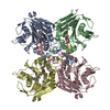 7svgC  7svhC 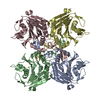 7sviC  7svjC  7svkC  2hezS S: Starting model for refinement C: citing same article ( |
|---|---|
| Similar structure data |
-Links
 PDBj
PDBj- Assembly
| Deposited unit | 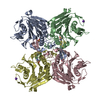
| ||||||||||||
|---|---|---|---|---|---|---|---|---|---|---|---|---|---|
| 1 |
| ||||||||||||
| Unit cell |
|
-Components
| #1: Protein | Mass: 35860.262 Da / Num. of mol.: 4 Source method: isolated from a genetically manipulated source Source: (gene. exp.) Lactobacillus gasseri (bacteria) / Gene: bsh, J3E66_000057 / Production host: #2: Chemical | ChemComp-TAU /   Mass: 125.147 Da / Num. of mol.: 4 / Source method: obtained synthetically / Formula: C2H7NO3S / Feature type: SUBJECT OF INVESTIGATION Mass: 125.147 Da / Num. of mol.: 4 / Source method: obtained synthetically / Formula: C2H7NO3S / Feature type: SUBJECT OF INVESTIGATION#3: Chemical |   Mass: 39.098 Da / Num. of mol.: 2 / Source method: obtained synthetically / Formula: K Mass: 39.098 Da / Num. of mol.: 2 / Source method: obtained synthetically / Formula: K#4: Water | ChemComp-HOH / |  Mass: 18.015 Da / Num. of mol.: 936 / Source method: isolated from a natural source / Formula: H2O Mass: 18.015 Da / Num. of mol.: 936 / Source method: isolated from a natural source / Formula: H2OHas ligand of interest | Y | |
|---|
-Experimental details
-Experiment
| Experiment | Method: X-RAY DIFFRACTION / Number of used crystals: 1 |
|---|
- Sample preparation
| Crystal | Density Matthews: 2.87 Å3/Da / Density % sol: 57.21 % |
|---|---|
| Crystal grow | Temperature: 293 K / Method: vapor diffusion, sitting drop Details: 0.2M Potassium Sulfate, 20% (w/v) PEG 3350. Crystals formed in a 1:2 protein (9.55 mg/mL) to mother liquor ratio. |
-Data collection
| Diffraction | Mean temperature: 100 K / Serial crystal experiment: N |
|---|---|
| Diffraction source | Source: SYNCHROTRON / Site: APS / Beamline: 23-ID-B / Wavelength: 1 Å |
| Detector | Type: DECTRIS EIGER X 16M / Detector: PIXEL / Date: Aug 11, 2020 |
| Radiation | Protocol: SINGLE WAVELENGTH / Monochromatic (M) / Laue (L): M / Scattering type: x-ray |
| Radiation wavelength | Wavelength: 1 Å / Relative weight: 1 |
| Reflection | Resolution: 2.05→45 Å / Num. obs: 100601 / % possible obs: 99.4 % / Redundancy: 1.9 % / CC1/2: 0.994 / CC star: 0.998 / Rmerge(I) obs: 0.067 / Rrim(I) all: 0.096 / Net I/σ(I): 6 |
| Reflection shell | Resolution: 2.05→2.12 Å / Redundancy: 1.9 % / Rmerge(I) obs: 0.296 / Mean I/σ(I) obs: 2.3 / Num. unique obs: 9994 / CC1/2: 0.81 / CC star: 0.946 / Rpim(I) all: 0.2958 / Rrim(I) all: 0.418 / % possible all: 99.4 |
- Processing
| Software |
| ||||||||||||||||||||||||
|---|---|---|---|---|---|---|---|---|---|---|---|---|---|---|---|---|---|---|---|---|---|---|---|---|---|
| Refinement | Method to determine structure: MOLECULAR REPLACEMENT Starting model: 2HEZ Resolution: 2.05→45 Å / Cross valid method: FREE R-VALUE Stereochemistry target values: GeoStd + Monomer Library + CDL v1.2
| ||||||||||||||||||||||||
| Displacement parameters | Biso mean: 24.73 Å2 | ||||||||||||||||||||||||
| Refinement step | Cycle: LAST / Resolution: 2.05→45 Å
| ||||||||||||||||||||||||
| Refine LS restraints |
|
