 Movie
Movie Controller

[English] 日本語
 Yorodumi
- PDB-8esl: Bile Salt Hydrolase from a Bacteroidales species with covalent in... -
+ Open data

- Basic information
| Entry | Database: PDB / ID: 8esl | ||||||
|---|---|---|---|---|---|---|---|
| Title | Bile Salt Hydrolase from a Bacteroidales species with covalent inhibitor bound | ||||||
 Components Components | Choloylglycine hydrolase | ||||||
 Keywords Keywords | HYDROLASE/HYDROLASE INHIBITOR / bile salt hydrolase / HYDROLASE-HYDROLASE INHIBITOR complex | ||||||
| Function / homology | Chem-WSR Function and homology information Function and homology information | ||||||
| Biological species |  Bacteroidales (bacteria) Bacteroidales (bacteria) | ||||||
| Method |  X-RAY DIFFRACTION / SYNCHROTRON / MOLECULAR REPLACEMENT / Resolution: 3.11 Å X-RAY DIFFRACTION / SYNCHROTRON / MOLECULAR REPLACEMENT / Resolution: 3.11 Å | ||||||
 Authors Authors | Walker, M.E. / Redinbo, M.R. | ||||||
| Funding support |  United States, 1items United States, 1items
| ||||||
 Citation Citation | Journal: To Be Published Title: Structural diversity of bile salt hydrolases reveals rationale for substrate selectivity Authors: Walker, M.E. / Redinbo, M.R. | ||||||
| History |
|
- Structure visualization
| Structure viewer | Molecule: MolmilJmol/JSmol |
|---|
- Downloads & links
-Download
| PDBx/mmCIF format | 8esl.cif.gz | 287.3 KB | Display | PDBx/mmCIF format |
|---|---|---|---|---|
| PDB format | pdb8esl.ent.gz | 185.8 KB | Display | PDB format |
| PDBx/mmJSON format | 8esl.json.gz | Tree view | PDBx/mmJSON format | |
| Others |  Other downloads Other downloads |
-Validation report
| Arichive directory | https://data.pdbj.org/pub/pdb/validation_reports/es/8eslftp://data.pdbj.org/pub/pdb/validation_reports/es/8esl | HTTPS FTP |
|---|
-Related structure data
| Related structure data |  8esgC  8esiC  8eteC  8etfC  8etkC  8ewtC  8faoC C: citing same article ( |
|---|---|
| Similar structure data |
-Links
 PDBj
PDBj- Assembly
| Deposited unit | 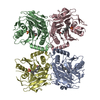
| ||||||||||||
|---|---|---|---|---|---|---|---|---|---|---|---|---|---|
| 1 |
| ||||||||||||
| Unit cell |
|
-Components
| #1: Protein | Mass: 37386.941 Da / Num. of mol.: 4 Source method: isolated from a genetically manipulated source Source: (gene. exp.) Bacteroidales (bacteria) / Production host: #2: Chemical | ChemComp-WSR / (   Mass: 472.653 Da / Num. of mol.: 4 / Source method: obtained synthetically / Formula: C25H41FO5S / Feature type: SUBJECT OF INVESTIGATION Mass: 472.653 Da / Num. of mol.: 4 / Source method: obtained synthetically / Formula: C25H41FO5S / Feature type: SUBJECT OF INVESTIGATIONHas ligand of interest | Y | Has protein modification | Y | |
|---|
-Experimental details
-Experiment
| Experiment | Method: X-RAY DIFFRACTION / Number of used crystals: 1 |
|---|
- Sample preparation
| Crystal | Density Matthews: 2.27 Å3/Da / Density % sol: 45.81 % |
|---|---|
| Crystal grow | Temperature: 293 K / Method: vapor diffusion, sitting drop Details: 0.1 M HEPES: NaOH, pH 7.5, 25% (w/v) PEG 1000. 50 mL protein at 2.5 uM was incubated with 50 uM inhibitor for 1h at 37oC. Mixture was washed 3x with buffer in a spin concentrator and then ...Details: 0.1 M HEPES: NaOH, pH 7.5, 25% (w/v) PEG 1000. 50 mL protein at 2.5 uM was incubated with 50 uM inhibitor for 1h at 37oC. Mixture was washed 3x with buffer in a spin concentrator and then concentrated to 6.77 mg/mL final concentration. Crystals formed in conditions with 1:2 ratio of protein:mother liquor. |
-Data collection
| Diffraction | Mean temperature: 100 K / Serial crystal experiment: N |
|---|---|
| Diffraction source | Source: SYNCHROTRON / Site: APS / Beamline: 23-ID-B / Wavelength: 1 Å |
| Detector | Type: DECTRIS EIGER X 16M / Detector: PIXEL / Date: Feb 27, 2022 |
| Radiation | Protocol: SINGLE WAVELENGTH / Monochromatic (M) / Laue (L): M / Scattering type: x-ray |
| Radiation wavelength | Wavelength: 1 Å / Relative weight: 1 |
| Reflection | Resolution: 3.11→45.07 Å / Num. obs: 24835 / % possible obs: 94.77 % / Redundancy: 3.4 % / Biso Wilson estimate: 92.36 Å2 / CC1/2: 0.978 / CC star: 0.994 / Rmerge(I) obs: 0.2399 / Rpim(I) all: 0.1551 / Rrim(I) all: 0.2875 / Net I/σ(I): 3.81 |
| Reflection shell | Resolution: 3.11→3.221 Å / Redundancy: 3.3 % / Rmerge(I) obs: 1.71 / Mean I/σ(I) obs: 0.74 / Num. unique obs: 2378 / CC1/2: 0.121 / CC star: 0.464 / Rpim(I) all: 1.115 / Rrim(I) all: 2.057 / % possible all: 80.39 |
- Processing
| Software |
| |||||||||||||||||||||||||||||||||||||||||||||||||||||||||||||||||||||||||||||||||||||||||||||||||||||||||
|---|---|---|---|---|---|---|---|---|---|---|---|---|---|---|---|---|---|---|---|---|---|---|---|---|---|---|---|---|---|---|---|---|---|---|---|---|---|---|---|---|---|---|---|---|---|---|---|---|---|---|---|---|---|---|---|---|---|---|---|---|---|---|---|---|---|---|---|---|---|---|---|---|---|---|---|---|---|---|---|---|---|---|---|---|---|---|---|---|---|---|---|---|---|---|---|---|---|---|---|---|---|---|---|---|---|---|
| Refinement | Method to determine structure: MOLECULAR REPLACEMENT Starting model: AlphaFold2 model of the protein Resolution: 3.11→45.07 Å / SU ML: 0.5378 / Cross valid method: FREE R-VALUE / σ(F): 1.33 / Phase error: 33.9647 Stereochemistry target values: GeoStd + Monomer Library + CDL v1.2
| |||||||||||||||||||||||||||||||||||||||||||||||||||||||||||||||||||||||||||||||||||||||||||||||||||||||||
| Solvent computation | Shrinkage radii: 0.9 Å / VDW probe radii: 1.1 Å / Solvent model: FLAT BULK SOLVENT MODEL | |||||||||||||||||||||||||||||||||||||||||||||||||||||||||||||||||||||||||||||||||||||||||||||||||||||||||
| Displacement parameters | Biso mean: 90.79 Å2 | |||||||||||||||||||||||||||||||||||||||||||||||||||||||||||||||||||||||||||||||||||||||||||||||||||||||||
| Refinement step | Cycle: LAST / Resolution: 3.11→45.07 Å
| |||||||||||||||||||||||||||||||||||||||||||||||||||||||||||||||||||||||||||||||||||||||||||||||||||||||||
| Refine LS restraints |
| |||||||||||||||||||||||||||||||||||||||||||||||||||||||||||||||||||||||||||||||||||||||||||||||||||||||||
| LS refinement shell |
|
