 Movie
Movie Controller


+ Open data

- Basic information
| Entry | Database: PDB / ID: 8e0h | |||||||||
|---|---|---|---|---|---|---|---|---|---|---|
| Title | Crystal structure of human Sar1aD104/D140A double mutant | |||||||||
 Components Components | GTP-binding protein SAR1a | |||||||||
 Keywords Keywords | SIGNALING PROTEIN / Sar1a / ppGpp / COPII / small GTPase | |||||||||
| Function / homology |  Function and homology information Function and homology informationamino acid sensor activity / : / regulation of TORC1 signaling / COPII-coated vesicle cargo loading / cellular response to leucine starvation / COPII vesicle coat assembly / vesicle organization / COPII vesicle coat / membrane organization / Golgi cisterna membrane ...amino acid sensor activity / : / regulation of TORC1 signaling / COPII-coated vesicle cargo loading / cellular response to leucine starvation / COPII vesicle coat assembly / vesicle organization / COPII vesicle coat / membrane organization / Golgi cisterna membrane / endoplasmic reticulum exit site / endoplasmic reticulum to Golgi vesicle-mediated transport / negative regulation of TORC1 signaling / small monomeric GTPase / intracellular protein transport / G protein activity / lysosomal membrane / GTPase activity / endoplasmic reticulum membrane / GTP binding / cytosol Similarity search - Function | |||||||||
| Biological species |  Homo sapiens (human) Homo sapiens (human) | |||||||||
| Method |  X-RAY DIFFRACTION / SYNCHROTRON / MOLECULAR REPLACEMENT / Resolution: 2 Å X-RAY DIFFRACTION / SYNCHROTRON / MOLECULAR REPLACEMENT / Resolution: 2 Å | |||||||||
 Authors Authors | Huang, Q. | |||||||||
| Funding support |  United States, 2items United States, 2items
| |||||||||
 Citation Citation | Journal: Proteins / Year: 2023 Title: The alarmone ppGpp selectively inhibits the isoform A of the human small GTPase Sar1. Authors: Huang, Q. / Szebenyi, D.M.E. | |||||||||
| History |
|
- Structure visualization
| Structure viewer | Molecule: MolmilJmol/JSmol |
|---|
- Downloads & links
-Download
| PDBx/mmCIF format | 8e0h.cif.gz | 158 KB | Display | PDBx/mmCIF format |
|---|---|---|---|---|
| PDB format | pdb8e0h.ent.gz | 122.5 KB | Display | PDB format |
| PDBx/mmJSON format | 8e0h.json.gz | Tree view | PDBx/mmJSON format | |
| Others |  Other downloads Other downloads |
-Validation report
| Arichive directory | https://data.pdbj.org/pub/pdb/validation_reports/e0/8e0hftp://data.pdbj.org/pub/pdb/validation_reports/e0/8e0h | HTTPS FTP |
|---|
-Related structure data
| Related structure data | 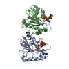 8dzmSC 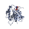 8dznC 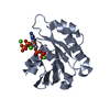 8dzoC 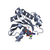 8dztC  8e0aC  8e0bC  8e0cC  8e0dC S: Starting model for refinement C: citing same article ( |
|---|---|
| Similar structure data |
-Links
 PDBj
PDBj


- Assembly
| Deposited unit | 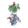
| ||||||||
|---|---|---|---|---|---|---|---|---|---|
| 1 | 
| ||||||||
| 2 | 
| ||||||||
| Unit cell |
|
-Components
| #1: Protein | Mass: 22304.754 Da / Num. of mol.: 2 / Mutation: D104A, D140A Source method: isolated from a genetically manipulated source Source: (gene. exp.) Homo sapiens (human) / Gene: SAR1A, SAR1, SARA, SARA1 / Production host:  #2: Chemical |   Type: RNA linking / Mass: 603.160 Da / Num. of mol.: 2 / Source method: obtained synthetically / Formula: C10H17N5O17P4 / Feature type: SUBJECT OF INVESTIGATION Type: RNA linking / Mass: 603.160 Da / Num. of mol.: 2 / Source method: obtained synthetically / Formula: C10H17N5O17P4 / Feature type: SUBJECT OF INVESTIGATION#3: Water | ChemComp-HOH / |  Mass: 18.015 Da / Num. of mol.: 166 / Source method: isolated from a natural source / Formula: H2O Mass: 18.015 Da / Num. of mol.: 166 / Source method: isolated from a natural source / Formula: H2OHas ligand of interest | Y | |
|---|
-Experimental details
-Experiment
| Experiment | Method: X-RAY DIFFRACTION / Number of used crystals: 1 |
|---|
- Sample preparation
| Crystal | Density Matthews: 2.04 Å3/Da / Density % sol: 39.76 % |
|---|---|
| Crystal grow | Temperature: 293 K / Method: vapor diffusion, hanging drop / Details: 3.5M NaCl, 0.2M CaCl2 and 0.1M NaAc, pH4.5. |
-Data collection
| Diffraction | Mean temperature: 100 K / Serial crystal experiment: N |
|---|---|
| Diffraction source | Source: SYNCHROTRON / Site: CHESS / Beamline: F1 / Wavelength: 0.9778 Å |
| Detector | Type: ADSC QUANTUM 270 / Detector: CCD / Date: May 12, 2018 |
| Radiation | Protocol: SINGLE WAVELENGTH / Monochromatic (M) / Laue (L): M / Scattering type: x-ray |
| Radiation wavelength | Wavelength: 0.9778 Å / Relative weight: 1 |
| Reflection | Resolution: 2→50 Å / Num. obs: 25484 / % possible obs: 99.9 % / Redundancy: 7.2 % / Rmerge(I) obs: 0.099 / Rpim(I) all: 0.039 / Rrim(I) all: 0.104 / Net I/σ(I): 32.3 |
| Reflection shell | Resolution: 2→2.03 Å / Redundancy: 6.7 % / Rmerge(I) obs: 0.361 / Mean I/σ(I) obs: 5.3 / Num. unique obs: 1262 / CC1/2: 0.945 / CC star: 0.986 / Rpim(I) all: 0.204 / Rrim(I) all: 0.537 / % possible all: 99.9 |
- Processing
| Software |
| |||||||||||||||||||||||||||||||||||||||||||||||||||||||||||||||||||||||||||||||||||||||||||||||||||||||||||||||||||||||||||||||||||||||||||||||||||||||||||||||||||||||||||||||||||||||||||||||||||||||||||||||||||||||||||||||||||||||||||||||||||||||||||||||||||||||||||||||||||||||||||||||||||||||||||||||||||||||||||||||||||||||||||||||||||||||||||||||||||||||||||||||||||||||
|---|---|---|---|---|---|---|---|---|---|---|---|---|---|---|---|---|---|---|---|---|---|---|---|---|---|---|---|---|---|---|---|---|---|---|---|---|---|---|---|---|---|---|---|---|---|---|---|---|---|---|---|---|---|---|---|---|---|---|---|---|---|---|---|---|---|---|---|---|---|---|---|---|---|---|---|---|---|---|---|---|---|---|---|---|---|---|---|---|---|---|---|---|---|---|---|---|---|---|---|---|---|---|---|---|---|---|---|---|---|---|---|---|---|---|---|---|---|---|---|---|---|---|---|---|---|---|---|---|---|---|---|---|---|---|---|---|---|---|---|---|---|---|---|---|---|---|---|---|---|---|---|---|---|---|---|---|---|---|---|---|---|---|---|---|---|---|---|---|---|---|---|---|---|---|---|---|---|---|---|---|---|---|---|---|---|---|---|---|---|---|---|---|---|---|---|---|---|---|---|---|---|---|---|---|---|---|---|---|---|---|---|---|---|---|---|---|---|---|---|---|---|---|---|---|---|---|---|---|---|---|---|---|---|---|---|---|---|---|---|---|---|---|---|---|---|---|---|---|---|---|---|---|---|---|---|---|---|---|---|---|---|---|---|---|---|---|---|---|---|---|---|---|---|---|---|---|---|---|---|---|---|---|---|---|---|---|---|---|---|---|---|---|---|---|---|---|---|---|---|---|---|---|---|---|---|---|---|---|---|---|---|---|---|---|---|---|---|---|---|---|---|---|---|---|---|---|---|---|---|---|---|---|---|---|---|---|---|---|---|---|---|---|---|---|---|---|---|---|---|---|---|---|---|---|---|---|---|---|---|---|---|---|---|---|---|---|---|---|---|---|---|---|---|---|---|---|
| Refinement | Method to determine structure: MOLECULAR REPLACEMENT Starting model: 8dzm Resolution: 2→45.131 Å / SU ML: 0.18 / Cross valid method: THROUGHOUT / σ(F): 1.33 / Phase error: 25.72 / Stereochemistry target values: ML
| |||||||||||||||||||||||||||||||||||||||||||||||||||||||||||||||||||||||||||||||||||||||||||||||||||||||||||||||||||||||||||||||||||||||||||||||||||||||||||||||||||||||||||||||||||||||||||||||||||||||||||||||||||||||||||||||||||||||||||||||||||||||||||||||||||||||||||||||||||||||||||||||||||||||||||||||||||||||||||||||||||||||||||||||||||||||||||||||||||||||||||||||||||||||
| Solvent computation | Shrinkage radii: 0.9 Å / VDW probe radii: 1.11 Å / Solvent model: FLAT BULK SOLVENT MODEL | |||||||||||||||||||||||||||||||||||||||||||||||||||||||||||||||||||||||||||||||||||||||||||||||||||||||||||||||||||||||||||||||||||||||||||||||||||||||||||||||||||||||||||||||||||||||||||||||||||||||||||||||||||||||||||||||||||||||||||||||||||||||||||||||||||||||||||||||||||||||||||||||||||||||||||||||||||||||||||||||||||||||||||||||||||||||||||||||||||||||||||||||||||||||
| Displacement parameters | Biso max: 153.1 Å2 / Biso mean: 44.1134 Å2 / Biso min: 21.3 Å2 | |||||||||||||||||||||||||||||||||||||||||||||||||||||||||||||||||||||||||||||||||||||||||||||||||||||||||||||||||||||||||||||||||||||||||||||||||||||||||||||||||||||||||||||||||||||||||||||||||||||||||||||||||||||||||||||||||||||||||||||||||||||||||||||||||||||||||||||||||||||||||||||||||||||||||||||||||||||||||||||||||||||||||||||||||||||||||||||||||||||||||||||||||||||||
| Refinement step | Cycle: final / Resolution: 2→45.131 Å
| |||||||||||||||||||||||||||||||||||||||||||||||||||||||||||||||||||||||||||||||||||||||||||||||||||||||||||||||||||||||||||||||||||||||||||||||||||||||||||||||||||||||||||||||||||||||||||||||||||||||||||||||||||||||||||||||||||||||||||||||||||||||||||||||||||||||||||||||||||||||||||||||||||||||||||||||||||||||||||||||||||||||||||||||||||||||||||||||||||||||||||||||||||||||
| Refine LS restraints |
| |||||||||||||||||||||||||||||||||||||||||||||||||||||||||||||||||||||||||||||||||||||||||||||||||||||||||||||||||||||||||||||||||||||||||||||||||||||||||||||||||||||||||||||||||||||||||||||||||||||||||||||||||||||||||||||||||||||||||||||||||||||||||||||||||||||||||||||||||||||||||||||||||||||||||||||||||||||||||||||||||||||||||||||||||||||||||||||||||||||||||||||||||||||||
| LS refinement shell | Refine-ID: X-RAY DIFFRACTION / Rfactor Rfree error: 0
| |||||||||||||||||||||||||||||||||||||||||||||||||||||||||||||||||||||||||||||||||||||||||||||||||||||||||||||||||||||||||||||||||||||||||||||||||||||||||||||||||||||||||||||||||||||||||||||||||||||||||||||||||||||||||||||||||||||||||||||||||||||||||||||||||||||||||||||||||||||||||||||||||||||||||||||||||||||||||||||||||||||||||||||||||||||||||||||||||||||||||||||||||||||||
| Refinement TLS params. | Method: refined / Refine-ID: X-RAY DIFFRACTION
| |||||||||||||||||||||||||||||||||||||||||||||||||||||||||||||||||||||||||||||||||||||||||||||||||||||||||||||||||||||||||||||||||||||||||||||||||||||||||||||||||||||||||||||||||||||||||||||||||||||||||||||||||||||||||||||||||||||||||||||||||||||||||||||||||||||||||||||||||||||||||||||||||||||||||||||||||||||||||||||||||||||||||||||||||||||||||||||||||||||||||||||||||||||||
| Refinement TLS group |
|