 Movie
Movie Controller

[English] 日本語
 Yorodumi
- PDB-8dw9: Crystal structure of neutralizing antibody D29 Fab in complex wit... -
+ Open data

- Basic information
| Entry | Database: PDB / ID: 8dw9 | ||||||
|---|---|---|---|---|---|---|---|
| Title | Crystal structure of neutralizing antibody D29 Fab in complex with SARS-CoV-2 spike receptor binding domain (RBD) | ||||||
 Components Components |
| ||||||
 Keywords Keywords | VIRAL PROTEIN/IMMUNE SYSTEM / COVID-19 / SarbecoVirus / WIV1 RBD / Viral protein / Spike glycoprotein / Receptor Binding Protein / Neutralizing antibody / potent / 10-40 / Fab / VIRAL PROTEIN-IMMUNE SYSTEM complex | ||||||
| Function / homology |  Function and homology information Function and homology informationsymbiont-mediated disruption of host tissue / Maturation of spike protein / Translation of Structural Proteins / Virion Assembly and Release / host cell surface / host extracellular region / symbiont-mediated-mediated suppression of host tetherin activity / Induction of Cell-Cell Fusion / structural constituent of virion / positive regulation of viral entry into host cell ...symbiont-mediated disruption of host tissue / Maturation of spike protein / Translation of Structural Proteins / Virion Assembly and Release / host cell surface / host extracellular region / symbiont-mediated-mediated suppression of host tetherin activity / Induction of Cell-Cell Fusion / structural constituent of virion / positive regulation of viral entry into host cell / membrane fusion / host cell endoplasmic reticulum-Golgi intermediate compartment membrane / Attachment and Entry / entry receptor-mediated virion attachment to host cell / receptor-mediated virion attachment to host cell / host cell surface receptor binding / symbiont-mediated suppression of host innate immune response / endocytosis involved in viral entry into host cell / receptor ligand activity / fusion of virus membrane with host plasma membrane / fusion of virus membrane with host endosome membrane / viral envelope / symbiont entry into host cell / virion attachment to host cell / host cell plasma membrane / SARS-CoV-2 activates/modulates innate and adaptive immune responses / virion membrane / membrane / identical protein binding / plasma membrane Similarity search - Function | ||||||
| Biological species |   Severe acute respiratory syndrome coronavirus 2 Severe acute respiratory syndrome coronavirus 2 Homo sapiens (human) Homo sapiens (human) | ||||||
| Method |  X-RAY DIFFRACTION / SYNCHROTRON / MOLECULAR REPLACEMENT / molecular replacement / Resolution: 4 Å X-RAY DIFFRACTION / SYNCHROTRON / MOLECULAR REPLACEMENT / molecular replacement / Resolution: 4 Å | ||||||
 Authors Authors | Reddem, E.R. / Shapiro, L. | ||||||
| Funding support |  China, 1items China, 1items
| ||||||
 Citation Citation | Journal: Emerg Microbes Infect / Year: 2023 Title: Structural insights into broadly neutralizing antibodies elicited by hybrid immunity against SARS-CoV-2. Authors: Mengxiao Luo / Biao Zhou / Eswar R Reddem / Bingjie Tang / Bohao Chen / Runhong Zhou / Hang Liu / Lihong Liu / Phinikoula S Katsamba / Ka-Kit Au / Hiu-On Man / Kelvin Kai-Wang To / Kwok-Yung ...Authors: Mengxiao Luo / Biao Zhou / Eswar R Reddem / Bingjie Tang / Bohao Chen / Runhong Zhou / Hang Liu / Lihong Liu / Phinikoula S Katsamba / Ka-Kit Au / Hiu-On Man / Kelvin Kai-Wang To / Kwok-Yung Yuen / Lawrence Shapiro / Shangyu Dang / David D Ho / Zhiwei Chen /  Abstract: Increasing spread by SARS-CoV-2 Omicron variants challenges existing vaccines and broadly reactive neutralizing antibodies (bNAbs) against COVID-19. Here we determine the diversity, potency, breadth ...Increasing spread by SARS-CoV-2 Omicron variants challenges existing vaccines and broadly reactive neutralizing antibodies (bNAbs) against COVID-19. Here we determine the diversity, potency, breadth and structural insights of bNAbs derived from memory B cells of BNT162b2-vaccinee after homogeneous Omicron BA.1 breakthrough infection. The infection activates diverse memory B cell clonotypes for generating potent class I/II and III bNAbs with new epitopes mapped to the receptor-binding domain (RBD). The top eight bNAbs neutralize wildtype and BA.1 potently but display divergent IgH/IgL sequences and neuralization profiles against other variants of concern (VOCs). Two of them (P2D9 and P3E6) belonging to class III NAbs display comparable potency against BA.4/BA.5, although structural analysis reveals distinct modes of action. P3E6 neutralizes all variants tested through a unique bivalent interaction with two RBDs. Our findings provide new insights into hybrid immunity on BNT162b2-induced diverse memory B cells in response to Omicron breakthrough infection for generating diverse bNAbs with distinct structural basis. | ||||||
| History |
|
- Structure visualization
| Structure viewer | Molecule: MolmilJmol/JSmol |
|---|
- Downloads & links
-Download
| PDBx/mmCIF format | 8dw9.cif.gz | 211.6 KB | Display | PDBx/mmCIF format |
|---|---|---|---|---|
| PDB format | pdb8dw9.ent.gz | 162.3 KB | Display | PDB format |
| PDBx/mmJSON format | 8dw9.json.gz | Tree view | PDBx/mmJSON format | |
| Others |  Other downloads Other downloads |
-Validation report
| Arichive directory | https://data.pdbj.org/pub/pdb/validation_reports/dw/8dw9ftp://data.pdbj.org/pub/pdb/validation_reports/dw/8dw9 | HTTPS FTP |
|---|
-Related structure data
| Related structure data |  7ykjC 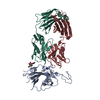 8dwaC  8dxsC 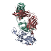 7sd5S S: Starting model for refinement C: citing same article ( |
|---|---|
| Similar structure data |


-Links
 PDBj
PDBj


- Assembly
| Deposited unit | 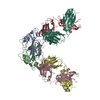
| ||||||||
|---|---|---|---|---|---|---|---|---|---|
| 1 | 
| ||||||||
| 2 | 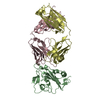
| ||||||||
| Unit cell |
|
-Components
| #1: Protein | Mass: 21873.496 Da / Num. of mol.: 2 / Fragment: receptor binding domain (UNP residues 333-527) Source method: isolated from a genetically manipulated source Source: (gene. exp.) Severe acute respiratory syndrome coronavirus 2Gene: S, 2 / Production host: Homo sapiens (human) / References: UniProt: P0DTC2#2: Antibody | Mass: 23284.070 Da / Num. of mol.: 2 Source method: isolated from a genetically manipulated source Source: (gene. exp.) Homo sapiens (human) / Production host: Homo sapiens (human)#3: Antibody | Mass: 22817.414 Da / Num. of mol.: 2 Source method: isolated from a genetically manipulated source Source: (gene. exp.) Homo sapiens (human) / Production host: Homo sapiens (human)Has protein modification | Y | |
|---|
-Experimental details
-Experiment
| Experiment | Method: X-RAY DIFFRACTION / Number of used crystals: 1 |
|---|
- Sample preparation
| Crystal | Density Matthews: 3.38 Å3/Da / Density % sol: 63.61 % |
|---|---|
| Crystal grow | Temperature: 293 K / Method: vapor diffusion, sitting drop Details: 2 M ammonium sulfate, 0.05 M MES, pH 6.0, 5 mM magnesium acetate tetrahydrate |
-Data collection
| Diffraction | Mean temperature: 100 K / Serial crystal experiment: N | ||||||||||||||||||||||||||||||
|---|---|---|---|---|---|---|---|---|---|---|---|---|---|---|---|---|---|---|---|---|---|---|---|---|---|---|---|---|---|---|---|
| Diffraction source | Source: SYNCHROTRON / Site: APS / Beamline: 24-ID-C / Wavelength: 0.97918 Å | ||||||||||||||||||||||||||||||
| Detector | Type: DECTRIS PILATUS 6M-F / Detector: PIXEL / Date: Mar 3, 2022 | ||||||||||||||||||||||||||||||
| Radiation | Protocol: SINGLE WAVELENGTH / Monochromatic (M) / Laue (L): M / Scattering type: x-ray | ||||||||||||||||||||||||||||||
| Radiation wavelength | Wavelength: 0.97918 Å / Relative weight: 1 | ||||||||||||||||||||||||||||||
| Reflection | Resolution: 3.25→146.18 Å / Num. obs: 15675 / % possible obs: 99.8 % / Redundancy: 7.4 % / CC1/2: 0.879 / Rmerge(I) obs: 0.957 / Rpim(I) all: 0.374 / Rrim(I) all: 1.029 / Net I/σ(I): 2.9 / Num. measured all: 416989 | ||||||||||||||||||||||||||||||
| Reflection shell | Diffraction-ID: 1
|
-Phasing
| Phasing | Method: molecular replacement |
|---|
- Processing
| Software |
| ||||||||||||||||||||
|---|---|---|---|---|---|---|---|---|---|---|---|---|---|---|---|---|---|---|---|---|---|
| Refinement | Method to determine structure: MOLECULAR REPLACEMENT Starting model: PDB entry 7SD5 Resolution: 4→95.09 Å / Cor.coef. Fo:Fc: 0.746 / Cor.coef. Fo:Fc free: 0.683 / SU B: 42 / SU ML: 0.6 / Cross valid method: THROUGHOUT / ESU R Free: 0.7 / Stereochemistry target values: MAXIMUM LIKELIHOOD Details: HYDROGENS HAVE BEEN ADDED IN THE RIDING POSITIONS U VALUES : REFINED INDIVIDUALLY
| ||||||||||||||||||||
| Solvent computation | Ion probe radii: 1.1 Å / Shrinkage radii: 1.1 Å / VDW probe radii: 1.2 Å / Solvent model: MASK | ||||||||||||||||||||
| Displacement parameters | Biso max: 41.47 Å2 / Biso mean: 40.213 Å2 / Biso min: 41.47 Å2
| ||||||||||||||||||||
| Refinement step | Cycle: final / Resolution: 4→95.09 Å
| ||||||||||||||||||||
| LS refinement shell | Resolution: 4→4.102 Å / Rfactor Rfree error: 0
|
