 Movie
Movie Controller


+ Open data

- Basic information
| Entry | Database: PDB / ID: 8d5b | ||||||||||||
|---|---|---|---|---|---|---|---|---|---|---|---|---|---|
| Title | Crystal structure of human METTL1 in complex with SAH | ||||||||||||
 Components Components | tRNA (guanine-N(7)-)-methyltransferase | ||||||||||||
 Keywords Keywords | TRANSFERASE / Cancer protein | ||||||||||||
| Function / homology |  Function and homology information Function and homology informationinternal mRNA (guanine-N7-)-methyltransferase activity / tRNA (m7G46) methyltransferase complex / tRNA (guanine-N7)-methylation / RNA (guanine-N7)-methylation / tRNA (guanine46-N7)-methyltransferase / tRNA (guanine(46)-N7)-methyltransferase activity / tRNA stabilization / tRNA methyltransferase complex / tRNA modification in the nucleus and cytosol / tRNA modification ...internal mRNA (guanine-N7-)-methyltransferase activity / tRNA (m7G46) methyltransferase complex / tRNA (guanine-N7)-methylation / RNA (guanine-N7)-methylation / tRNA (guanine46-N7)-methyltransferase / tRNA (guanine(46)-N7)-methyltransferase activity / tRNA stabilization / tRNA methyltransferase complex / tRNA modification in the nucleus and cytosol / tRNA modification / tRNA methylation / cellular response to stress / Transferases; Transferring one-carbon groups; Methyltransferases / tRNA binding / nucleolus / nucleoplasm / nucleus / cytosol Similarity search - Function | ||||||||||||
| Biological species |  Homo sapiens (human) Homo sapiens (human) | ||||||||||||
| Method |  X-RAY DIFFRACTION / SYNCHROTRON / MOLECULAR REPLACEMENT / Resolution: 1.93 Å X-RAY DIFFRACTION / SYNCHROTRON / MOLECULAR REPLACEMENT / Resolution: 1.93 Å | ||||||||||||
 Authors Authors | Raj, R. / Babu, K. / Nam, Y. | ||||||||||||
| Funding support |  United States, 3items United States, 3items
| ||||||||||||
 Citation Citation | Journal: Nature / Year: 2023 Title: Structures and mechanisms of tRNA methylation by METTL1-WDR4. Authors: Victor M Ruiz-Arroyo / Rishi Raj / Kesavan Babu / Otgonbileg Onolbaatar / Paul H Roberts / Yunsun Nam / Abstract: Specific, regulated modification of RNAs is important for proper gene expression. tRNAs are rich with various chemical modifications that affect their stability and function. 7-Methylguanosine (mG) ...Specific, regulated modification of RNAs is important for proper gene expression. tRNAs are rich with various chemical modifications that affect their stability and function. 7-Methylguanosine (mG) at tRNA position 46 is a conserved modification that modulates steady-state tRNA levels to affect cell growth. The METTL1-WDR4 complex generates mG46 in humans, and dysregulation of METTL1-WDR4 has been linked to brain malformation and multiple cancers. Here we show how METTL1 and WDR4 cooperate to recognize RNA substrates and catalyse methylation. A crystal structure of METTL1-WDR4 and cryo-electron microscopy structures of METTL1-WDR4-tRNA show that the composite protein surface recognizes the tRNA elbow through shape complementarity. The cryo-electron microscopy structures of METTL1-WDR4-tRNA with S-adenosylmethionine or S-adenosylhomocysteine along with METTL1 crystal structures provide additional insights into the catalytic mechanism by revealing the active site in multiple states. The METTL1 N terminus couples cofactor binding with conformational changes in the tRNA, the catalytic loop and the WDR4 C terminus, acting as the switch to activate mG methylation. Thus, our structural models explain how post-translational modifications of the METTL1 N terminus can regulate methylation. Together, our work elucidates the core and regulatory mechanisms underlying mG modification by METTL1, providing the framework to understand its contribution to biology and disease. | ||||||||||||
| History |
|
- Structure visualization
| Structure viewer | Molecule: MolmilJmol/JSmol |
|---|
- Downloads & links
-Download
| PDBx/mmCIF format | 8d5b.cif.gz | 74.3 KB | Display | PDBx/mmCIF format |
|---|---|---|---|---|
| PDB format | pdb8d5b.ent.gz | 41.8 KB | Display | PDB format |
| PDBx/mmJSON format | 8d5b.json.gz | Tree view | PDBx/mmJSON format | |
| Others |  Other downloads Other downloads |
-Validation report
| Arichive directory | https://data.pdbj.org/pub/pdb/validation_reports/d5/8d5bftp://data.pdbj.org/pub/pdb/validation_reports/d5/8d5b | HTTPS FTP |
|---|
-Related structure data
| Related structure data | 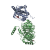 8d58C 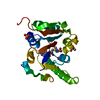 8d59C  8d9kC 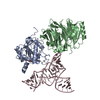 8d9lC 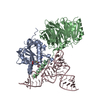 8eg0C 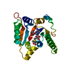 3ckkS S: Starting model for refinement C: citing same article ( |
|---|---|
| Similar structure data |
-Links
 PDBj
PDBj


- Assembly
| Deposited unit | 
| ||||||||||||
|---|---|---|---|---|---|---|---|---|---|---|---|---|---|
| 1 |
| ||||||||||||
| Unit cell |
|
-Components
-Protein , 1 types, 1 molecules A
| #1: Protein | Mass: 30412.713 Da / Num. of mol.: 1 Source method: isolated from a genetically manipulated source Source: (gene. exp.) Homo sapiens (human) / Gene: METTL1, C12orf1 / Production host:  References: UniProt: Q9UBP6, tRNA (guanine46-N7)-methyltransferase, Transferases; Transferring one-carbon groups; Methyltransferases |
|---|
-Non-polymers , 6 types, 151 molecules 


| #2: Chemical | ChemComp-SAH /  Type: L-peptide linking / Mass: 384.411 Da / Num. of mol.: 1 / Source method: obtained synthetically / Formula: C14H20N6O5S / Feature type: SUBJECT OF INVESTIGATION Type: L-peptide linking / Mass: 384.411 Da / Num. of mol.: 1 / Source method: obtained synthetically / Formula: C14H20N6O5S / Feature type: SUBJECT OF INVESTIGATION | ||||||||
|---|---|---|---|---|---|---|---|---|---|
| #3: Chemical |  Mass: 96.063 Da / Num. of mol.: 2 / Source method: obtained synthetically / Formula: SO4 Mass: 96.063 Da / Num. of mol.: 2 / Source method: obtained synthetically / Formula: SO4#4: Chemical |  Mass: 92.094 Da / Num. of mol.: 2 / Source method: isolated from a natural source / Formula: C3H8O3 Mass: 92.094 Da / Num. of mol.: 2 / Source method: isolated from a natural source / Formula: C3H8O3#5: Chemical | ChemComp-CL / |  Mass: 35.453 Da / Num. of mol.: 1 / Source method: obtained synthetically / Formula: Cl Mass: 35.453 Da / Num. of mol.: 1 / Source method: obtained synthetically / Formula: Cl#6: Chemical | ChemComp-CAC / |  Mass: 136.989 Da / Num. of mol.: 1 / Source method: isolated from a natural source / Formula: C2H6AsO2 Mass: 136.989 Da / Num. of mol.: 1 / Source method: isolated from a natural source / Formula: C2H6AsO2#7: Water | ChemComp-HOH / | Mass: 18.015 Da / Num. of mol.: 144 / Source method: isolated from a natural source / Formula: H2O |
-Details
| Has ligand of interest | Y |
|---|
-Experimental details
-Experiment
| Experiment | Method: X-RAY DIFFRACTION / Number of used crystals: 1 |
|---|
- Sample preparation
| Crystal | Density Matthews: 2.46 Å3/Da / Density % sol: 49.97 % |
|---|---|
| Crystal grow | Temperature: 295.15 K / Method: vapor diffusion, hanging drop Details: 50 mM Sodium cacodylate pH 5.7, 1.5 M Lithium sulfate, 10 mM Magnesium sulfate |
-Data collection
| Diffraction | Mean temperature: 100 K / Serial crystal experiment: N |
|---|---|
| Diffraction source | Source: SYNCHROTRON / Site: APS / Beamline: 19-ID / Wavelength: 0.9789 Å |
| Detector | Type: DECTRIS PILATUS3 6M / Detector: PIXEL / Date: Dec 2, 2020 |
| Radiation | Protocol: SINGLE WAVELENGTH / Monochromatic (M) / Laue (L): M / Scattering type: x-ray |
| Radiation wavelength | Wavelength: 0.9789 Å / Relative weight: 1 |
| Reflection | Resolution: 1.93→37.25 Å / Num. obs: 20856 / % possible obs: 100 % / Redundancy: 18.1 % / Biso Wilson estimate: 32.6 Å2 / CC1/2: 0.996 / Rmerge(I) obs: 0.242 / Rpim(I) all: 0.057 / Net I/σ(I): 16.24 |
| Reflection shell | Resolution: 1.93→1.96 Å / Redundancy: 10.5 % / Rmerge(I) obs: 3.22 / Mean I/σ(I) obs: 1.3 / Num. unique obs: 1034 / CC1/2: 0.45 / Rpim(I) all: 1.03 / % possible all: 99.9 |
- Processing
| Software |
| ||||||||||||||||||||||||||||||||||||||||||||||||||||||||
|---|---|---|---|---|---|---|---|---|---|---|---|---|---|---|---|---|---|---|---|---|---|---|---|---|---|---|---|---|---|---|---|---|---|---|---|---|---|---|---|---|---|---|---|---|---|---|---|---|---|---|---|---|---|---|---|---|---|
| Refinement | Method to determine structure: MOLECULAR REPLACEMENT Starting model: 3CKK Resolution: 1.93→37.25 Å / SU ML: 0.2564 / Cross valid method: FREE R-VALUE / σ(F): 1.35 / Phase error: 24.2094 Stereochemistry target values: GeoStd + Monomer Library + CDL v1.2
| ||||||||||||||||||||||||||||||||||||||||||||||||||||||||
| Solvent computation | Shrinkage radii: 0.9 Å / VDW probe radii: 1.1 Å / Solvent model: FLAT BULK SOLVENT MODEL | ||||||||||||||||||||||||||||||||||||||||||||||||||||||||
| Displacement parameters | Biso mean: 36.9 Å2 | ||||||||||||||||||||||||||||||||||||||||||||||||||||||||
| Refinement step | Cycle: LAST / Resolution: 1.93→37.25 Å
| ||||||||||||||||||||||||||||||||||||||||||||||||||||||||
| Refine LS restraints |
| ||||||||||||||||||||||||||||||||||||||||||||||||||||||||
| LS refinement shell |
|
