 Movie
Movie Controller

[English] 日本語
 Yorodumi
- PDB-8cvo: Cutibacterium acnes 30S ribosomal subunit with Sarecycline bound,... -
+ Open data

- Basic information
| Entry | Database: PDB / ID: 8cvo | ||||||
|---|---|---|---|---|---|---|---|
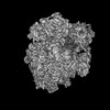
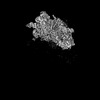
| Title | Cutibacterium acnes 30S ribosomal subunit with Sarecycline bound, head domain only in the local refined map | ||||||
 Components Components |
| ||||||
 Keywords Keywords | ANTIBIOTIC / ribosome / sarecycline / acne | ||||||
| Function / homology |  Function and homology information Function and homology informationribosomal small subunit assembly / ribosome biogenesis / small ribosomal subunit / cytosolic small ribosomal subunit / tRNA binding / rRNA binding / structural constituent of ribosome / ribosome / translation / ribonucleoprotein complex ...ribosomal small subunit assembly / ribosome biogenesis / small ribosomal subunit / cytosolic small ribosomal subunit / tRNA binding / rRNA binding / structural constituent of ribosome / ribosome / translation / ribonucleoprotein complex / mRNA binding / RNA binding / zinc ion binding / cytosol / cytoplasm Similarity search - Function | ||||||
| Biological species |  Cutibacterium acnes (bacteria) Cutibacterium acnes (bacteria) | ||||||
| Method | ELECTRON MICROSCOPY / single particle reconstruction / cryo EM / Resolution: 2.95 Å | ||||||
 Authors Authors | Lomakin, I.B. / Devarkar, S.C. / Bunick, C.G. | ||||||
| Funding support |  United States, 1items United States, 1items
| ||||||
 Citation Citation | Journal: Nucleic Acids Res / Year: 2023 Title: Sarecycline inhibits protein translation in Cutibacterium acnes 70S ribosome using a two-site mechanism. Authors: Ivan B Lomakin / Swapnil C Devarkar / Shivali Patel / Ayman Grada / Christopher G Bunick / Abstract: Acne vulgaris is a chronic disfiguring skin disease affecting ∼1 billion people worldwide, often having persistent negative effects on physical and mental health. The Gram-positive anaerobe, ...Acne vulgaris is a chronic disfiguring skin disease affecting ∼1 billion people worldwide, often having persistent negative effects on physical and mental health. The Gram-positive anaerobe, Cutibacterium acnes is implicated in acne pathogenesis and is, therefore, a main target for antibiotic-based acne therapy. We determined a 2.8-Å resolution structure of the 70S ribosome of Cutibacterium acnes by cryogenic electron microscopy and discovered that sarecycline, a narrow-spectrum antibiotic against Cutibacterium acnes, may inhibit two active sites of this bacterium's ribosome in contrast to the one site detected previously on the model ribosome of Thermus thermophilus. Apart from the canonical binding site at the mRNA decoding center, the second binding site for sarecycline exists at the nascent peptide exit tunnel, reminiscent of the macrolides class of antibiotics. The structure also revealed Cutibacterium acnes-specific features of the ribosomal RNA and proteins. Unlike the ribosome of the Gram-negative bacterium Escherichia coli, Cutibacterium acnes ribosome has two additional proteins, bS22 and bL37, which are also present in the ribosomes of Mycobacterium smegmatis and Mycobacterium tuberculosis. We show that bS22 and bL37 have antimicrobial properties and may be involved in maintaining the healthy homeostasis of the human skin microbiome. #1: Journal: To be publishedTitle: The structure of the Cutibacterium acnes 70S ribosome Authors: Lomakin, I.B. / Devarkar, S.C. / Grada, A. / Bunick, C.G. | ||||||
| History |
|
- Structure visualization
| Structure viewer | Molecule: MolmilJmol/JSmol |
|---|
- Downloads & links
-Download
| PDBx/mmCIF format | 8cvo.cif.gz | 431.6 KB | Display | PDBx/mmCIF format |
|---|---|---|---|---|
| PDB format | pdb8cvo.ent.gz | 314.4 KB | Display | PDB format |
| PDBx/mmJSON format | 8cvo.json.gz | Tree view | PDBx/mmJSON format | |
| Others |  Other downloads Other downloads |
-Validation report
| Arichive directory | https://data.pdbj.org/pub/pdb/validation_reports/cv/8cvoftp://data.pdbj.org/pub/pdb/validation_reports/cv/8cvo | HTTPS FTP |
|---|
-Related structure data
| Related structure data |  27011MC  8crxC 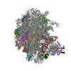 8cvmC  8cwoC C: citing same article ( M: map data used to model this data |
|---|---|
| Similar structure data |


-Links
 PDBj
PDBj


- Assembly
| Deposited unit | 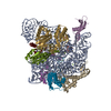
|
|---|---|
| 1 |
|
-Components
-RNA chain , 1 types, 1 molecules A
| #1: RNA chain | Mass: 498742.875 Da / Num. of mol.: 1 / Source method: isolated from a natural source / Source: (natural) Cutibacterium acnes (bacteria) / References: GenBank: 2179609306 |
|---|
-30S ribosomal protein ... , 8 types, 8 molecules EGIJMNSU
| #2: Protein | Mass: 22603.695 Da / Num. of mol.: 1 / Source method: isolated from a natural source / Source: (natural) Cutibacterium acnes (bacteria) / References: UniProt: A0A2B7I5L8 |
|---|---|
| #3: Protein | Mass: 29728.719 Da / Num. of mol.: 1 / Source method: isolated from a natural source / Source: (natural) Cutibacterium acnes (bacteria) / References: UniProt: A0A2B7I5Y3 |
| #4: Protein | Mass: 17615.482 Da / Num. of mol.: 1 / Source method: isolated from a natural source / Source: (natural) Cutibacterium acnes (bacteria) / References: UniProt: A0A2B7ITZ4 |
| #5: Protein | Mass: 18630.139 Da / Num. of mol.: 1 / Source method: isolated from a natural source / Source: (natural) Cutibacterium acnes (bacteria) / References: UniProt: A0A2B7JN42 |
| #6: Protein | Mass: 11750.766 Da / Num. of mol.: 1 / Source method: isolated from a natural source / Source: (natural) Cutibacterium acnes (bacteria) / References: UniProt: A0A085B5E4 |
| #7: Protein | Mass: 14030.231 Da / Num. of mol.: 1 / Source method: isolated from a natural source / Source: (natural) Cutibacterium acnes (bacteria) / References: UniProt: A0A2C6LKT6 |
| #8: Protein | Mass: 6924.359 Da / Num. of mol.: 1 / Source method: isolated from a natural source / Source: (natural) Cutibacterium acnes (bacteria) / References: UniProt: A0A2B7JMX1 |
| #9: Protein | Mass: 10536.264 Da / Num. of mol.: 1 / Source method: isolated from a natural source / Source: (natural) Cutibacterium acnes (bacteria) / References: UniProt: A0A2B7ITR7 |
-Non-polymers , 3 types, 14 molecules 


| #10: Chemical | ChemComp-V7A /  Mass: 487.502 Da / Num. of mol.: 1 / Source method: obtained synthetically / Formula: C24H29N3O8 / Feature type: SUBJECT OF INVESTIGATION / Comment: medication, antibiotic*YM Mass: 487.502 Da / Num. of mol.: 1 / Source method: obtained synthetically / Formula: C24H29N3O8 / Feature type: SUBJECT OF INVESTIGATION / Comment: medication, antibiotic*YM | ||
|---|---|---|---|
| #11: Chemical | ChemComp-MG /  Mass: 24.305 Da / Num. of mol.: 12 / Source method: obtained synthetically / Formula: Mg Mass: 24.305 Da / Num. of mol.: 12 / Source method: obtained synthetically / Formula: Mg#12: Chemical | ChemComp-ZN / |  Mass: 65.409 Da / Num. of mol.: 1 / Source method: obtained synthetically / Formula: Zn Mass: 65.409 Da / Num. of mol.: 1 / Source method: obtained synthetically / Formula: Zn |
-Details
| Has ligand of interest | Y |
|---|
-Experimental details
-Experiment
| Experiment | Method: ELECTRON MICROSCOPY |
|---|---|
| EM experiment | Aggregation state: PARTICLE / 3D reconstruction method: single particle reconstruction |
- Sample preparation
| Component | Name: 30S subunit head domain from 70S ribosome with mRNA, P-site tRNA and Sarecycline bound Type: RIBOSOME / Entity ID: #1-#9 / Source: NATURAL |
|---|---|
| Molecular weight | Experimental value: NO |
| Source (natural) | Organism: Cutibacterium acnes (bacteria) |
| Buffer solution | pH: 7.4 |
| Specimen | Embedding applied: NO / Shadowing applied: NO / Staining applied: NO / Vitrification applied: YES |
| Vitrification | Instrument: FEI VITROBOT MARK IV / Cryogen name: ETHANE / Humidity: 100 % |
- Electron microscopy imaging
| Experimental equipment |  Model: Titan Krios / Image courtesy: FEI Company |
|---|---|
| Microscopy | Model: FEI TITAN KRIOS |
| Electron gun | Electron source:  FIELD EMISSION GUN / Accelerating voltage: 300 kV / Illumination mode: FLOOD BEAM FIELD EMISSION GUN / Accelerating voltage: 300 kV / Illumination mode: FLOOD BEAM |
| Electron lens | Mode: BRIGHT FIELD / Nominal defocus max: 2000 nm / Nominal defocus min: 500 nm / Cs: 2.7 mm / C2 aperture diameter: 50 µm |
| Specimen holder | Cryogen: NITROGEN |
| Image recording | Average exposure time: 2.1 sec. / Electron dose: 30.56 e/Å2 / Film or detector model: GATAN K3 (6k x 4k) |
- Processing
| EM software |
| |||||||||||||||||||||||||||||||||||||||||||||
|---|---|---|---|---|---|---|---|---|---|---|---|---|---|---|---|---|---|---|---|---|---|---|---|---|---|---|---|---|---|---|---|---|---|---|---|---|---|---|---|---|---|---|---|---|---|---|
| CTF correction | Type: PHASE FLIPPING AND AMPLITUDE CORRECTION | |||||||||||||||||||||||||||||||||||||||||||||
| Particle selection | Num. of particles selected: 1108852 | |||||||||||||||||||||||||||||||||||||||||||||
| 3D reconstruction | Resolution: 2.95 Å / Resolution method: FSC 0.143 CUT-OFF / Num. of particles: 70853 / Symmetry type: POINT | |||||||||||||||||||||||||||||||||||||||||||||
| Atomic model building | Protocol: FLEXIBLE FIT / Space: REAL |
