purine-rich negative regulatory element binding / dendritic transport of messenger ribonucleoprotein complex / lymphocyte proliferation / double-stranded telomeric DNA binding / SMAD binding / DNA replication initiation / transcription regulator inhibitor activity / mRNA regulatory element binding translation repressor activity / epithelial cell proliferation / RNA polymerase II transcription regulatory region sequence-specific DNA binding ...purine-rich negative regulatory element binding / dendritic transport of messenger ribonucleoprotein complex / lymphocyte proliferation / double-stranded telomeric DNA binding / SMAD binding / DNA replication initiation / transcription regulator inhibitor activity / mRNA regulatory element binding translation repressor activity / epithelial cell proliferation / RNA polymerase II transcription regulatory region sequence-specific DNA binding / DNA-binding transcription repressor activity, RNA polymerase II-specific / nervous system development / single-stranded DNA binding / DNA-binding transcription factor binding / DNA-binding transcription factor activity, RNA polymerase II-specific / chromosome, telomeric region / postsynapse / neuronal cell body / positive regulation of cell population proliferation / dendrite / regulation of transcription by RNA polymerase II / glutamatergic synapse / negative regulation of transcription by RNA polymerase II / RNA binding / nucleus / cytoplasm Similarity search - Function
PurA ssDNA and RNA-binding protein / Purine-rich element binding protein family / DNA/RNA-binding repeats in PUR-alpha/beta/gamma and in hypothetical proteins from spirochetes and the Bacteroides-Cytophaga-Flexibacter bacteria. Similarity search - Domain/homology
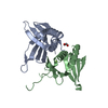
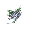
A: Transcriptional activator protein Pur-alpha B: Transcriptional activator protein Pur-alpha C: Transcriptional activator protein Pur-alpha D: Transcriptional activator protein Pur-alpha
Protocol: SINGLE WAVELENGTH / Monochromatic (M) / Laue (L): M / Scattering type: x-ray
Radiation wavelength
Wavelength: 1 Å / Relative weight: 1
Reflection
Resolution: 2.15→50 Å / Num. obs: 32410 / % possible obs: 99.4 % / Redundancy: 3.5 % / CC1/2: 0.997 / Net I/σ(I): 7.6
Reflection shell
Resolution: 2.15→2.28 Å / Num. unique obs: 5012 / CC1/2: 0.583 / % possible all: 97.7
-
Processing
Software
Name
Version
Classification
REFMAC
5.8.0352
refinement
PDB_EXTRACT
3.27
dataextraction
XDS
datareduction
SCALA
datascaling
MOLREP
phasing
Refinement
Method to determine structure: MOLECULAR REPLACEMENT / Resolution: 2.15→47.29 Å / Cor.coef. Fo:Fc: 0.955 / Cor.coef. Fo:Fc free: 0.917 / SU B: 20.086 / SU ML: 0.241 / Cross valid method: THROUGHOUT / σ(F): 0 / ESU R: 0.289 / ESU R Free: 0.244 / Stereochemistry target values: MAXIMUM LIKELIHOOD Details: HYDROGENS HAVE BEEN ADDED IN THE RIDING POSITIONS U VALUES : WITH TLS ADDED
Rfactor
Num. reflection
% reflection
Selection details
Rfree
0.2821
1621
5 %
RANDOM
Rwork
0.2048
-
-
-
obs
0.2087
30789
99.37 %
-
Solvent computation
Ion probe radii: 0.8 Å / Shrinkage radii: 0.8 Å / VDW probe radii: 1.2 Å / Solvent model: MASK
In the structure databanks used in Yorodumi, some data are registered as the other names, "COVID-19 virus" and "2019-nCoV". Here are the details of the virus and the list of structure data.
Jan 31, 2019. EMDB accession codes are about to change! (news from PDBe EMDB page)
EMDB accession codes are about to change! (news from PDBe EMDB page)
The allocation of 4 digits for EMDB accession codes will soon come to an end. Whilst these codes will remain in use, new EMDB accession codes will include an additional digit and will expand incrementally as the available range of codes is exhausted. The current 4-digit format prefixed with “EMD-” (i.e. EMD-XXXX) will advance to a 5-digit format (i.e. EMD-XXXXX), and so on. It is currently estimated that the 4-digit codes will be depleted around Spring 2019, at which point the 5-digit format will come into force.
The EM Navigator/Yorodumi systems omit the EMD- prefix.
Related info.:Q: What is EMD? / ID/Accession-code notation in Yorodumi/EM Navigator
Yorodumi is a browser for structure data from EMDB, PDB, SASBDB, etc.
This page is also the successor to EM Navigator detail page, and also detail information page/front-end page for Omokage search.
The word "yorodu" (or yorozu) is an old Japanese word meaning "ten thousand". "mi" (miru) is to see.
Related info.:EMDB / PDB / SASBDB / Comparison of 3 databanks / Yorodumi Search / Aug 31, 2016. New EM Navigator & Yorodumi / Yorodumi Papers / Jmol/JSmol / Function and homology information / Changes in new EM Navigator and Yorodumi
 Movie
Movie Controller

 Yorodumi
 Open data
 Basic information
 Components
 Keywords
 Function and homology information
 Homo sapiens (human)
 X-RAY DIFFRACTION /
  Authors
 Germany, 1items
  Citation
 Structure visualization
 Downloads & links
 Other downloads
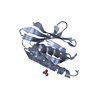
 PDBj
PDBj Assembly
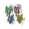

 Mass: 18.015 Da / Num. of mol.: 197 / Source method: isolated from a natural source / Formula: H2O
 Sample preparation
 / Beamline: X06DA / Wavelength: 1 Å
 Processing