 Movie
Movie Controller


+ Open data

- Basic information
| Entry | Database: PDB / ID: 8btx | ||||||
|---|---|---|---|---|---|---|---|
| Title | Structure of human Archease | ||||||
 Components Components | Protein archease | ||||||
 Keywords Keywords | LIGASE / archease / human / tRNA splicing | ||||||
| Function / homology | Archease / Archease domain / Archease domain superfamily / Archease protein family (MTH1598/TM1083) / tRNA processing / metal ion binding / Protein archease Function and homology information Function and homology information | ||||||
| Biological species |  Homo sapiens (human) Homo sapiens (human) | ||||||
| Method |  X-RAY DIFFRACTION / SYNCHROTRON / MOLECULAR REPLACEMENT / Resolution: 1.84 Å X-RAY DIFFRACTION / SYNCHROTRON / MOLECULAR REPLACEMENT / Resolution: 1.84 Å | ||||||
 Authors Authors | Kopp, J. / Gerber, J.L. / Peschek, J. | ||||||
| Funding support |  Germany, 1items Germany, 1items
| ||||||
 Citation Citation | Journal: Nat Commun / Year: 2024 Title: Structural and mechanistic insights into activation of the human RNA ligase RTCB by Archease. Authors: Gerber, J.L. / Morales Guzman, S.I. / Worf, L. / Hubbe, P. / Kopp, J. / Peschek, J. | ||||||
| History |
|
- Structure visualization
| Structure viewer | Molecule: MolmilJmol/JSmol |
|---|
- Downloads & links
-Download
| PDBx/mmCIF format | 8btx.cif.gz | 95.2 KB | Display | PDBx/mmCIF format |
|---|---|---|---|---|
| PDB format | pdb8btx.ent.gz | 60.2 KB | Display | PDB format |
| PDBx/mmJSON format | 8btx.json.gz | Tree view | PDBx/mmJSON format | |
| Others |  Other downloads Other downloads |
-Validation report
| Arichive directory | https://data.pdbj.org/pub/pdb/validation_reports/bt/8btxftp://data.pdbj.org/pub/pdb/validation_reports/bt/8btx | HTTPS FTP |
|---|
-Related structure data
| Related structure data |  8bttC 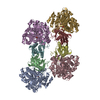 8odoC  8odpC 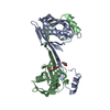 5yz1S C: citing same article ( S: Starting model for refinement |
|---|---|
| Similar structure data |
-Links
 PDBj
PDBj- Assembly
| Deposited unit | 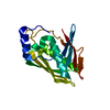
| ||||||||||||
|---|---|---|---|---|---|---|---|---|---|---|---|---|---|
| 1 |
| ||||||||||||
| Unit cell |
|
-Components
| #1: Protein | Mass: 21108.562 Da / Num. of mol.: 1 Source method: isolated from a genetically manipulated source Source: (gene. exp.) Homo sapiens (human) / Gene: ZBTB8OS / Production host:  |
|---|---|
| #2: Water | ChemComp-HOH /  Mass: 18.015 Da / Num. of mol.: 43 / Source method: isolated from a natural source / Formula: H2O Mass: 18.015 Da / Num. of mol.: 43 / Source method: isolated from a natural source / Formula: H2O |
-Experimental details
-Experiment
| Experiment | Method: X-RAY DIFFRACTION / Number of used crystals: 1 |
|---|
- Sample preparation
| Crystal | Density Matthews: 2.47 Å3/Da / Density % sol: 50.17 % |
|---|---|
| Crystal grow | Temperature: 291.15 K / Method: vapor diffusion, sitting drop Details: protein: 1966 uM in 25 mM HEPES pH 7.5, 100mM NaCl, 5 % glycerol, 1mM TCEP reservoir: 0.2 M ammonium chloride, 20% w/v PEG 3350 drop composition: 200 nl reservoir + 200 nl protein |
-Data collection
| Diffraction | Mean temperature: 100 K / Serial crystal experiment: N |
|---|---|
| Diffraction source | Source: SYNCHROTRON / Site: ESRF  / Beamline: ID23-1 / Wavelength: 0.9737 Å / Beamline: ID23-1 / Wavelength: 0.9737 Å |
| Detector | Type: DECTRIS EIGER2 X 16M / Detector: PIXEL / Date: Sep 3, 2021 / Details: Toroidal mirror |
| Radiation | Monochromator: Si(111) / Protocol: SINGLE WAVELENGTH / Monochromatic (M) / Laue (L): M / Scattering type: x-ray |
| Radiation wavelength | Wavelength: 0.9737 Å / Relative weight: 1 |
| Reflection | Resolution: 1.84→79.98 Å / Num. obs: 18949 / % possible obs: 100 % / Redundancy: 27.5 % / Biso Wilson estimate: 49.91 Å2 / CC1/2: 0.999 / Rmerge(I) obs: 0.068 / Rpim(I) all: 0.013 / Net I/σ(I): 23.7 |
| Reflection shell | Resolution: 1.84→1.88 Å / Redundancy: 25.1 % / Num. unique obs: 1137 / CC1/2: 0.471 / Rpim(I) all: 0.743 / % possible all: 100 |
- Processing
| Software |
| |||||||||||||||||||||||||||||||||||||||||||||||||||||||||||||||||||||||||||
|---|---|---|---|---|---|---|---|---|---|---|---|---|---|---|---|---|---|---|---|---|---|---|---|---|---|---|---|---|---|---|---|---|---|---|---|---|---|---|---|---|---|---|---|---|---|---|---|---|---|---|---|---|---|---|---|---|---|---|---|---|---|---|---|---|---|---|---|---|---|---|---|---|---|---|---|---|
| Refinement | Method to determine structure: MOLECULAR REPLACEMENT Starting model: 5YZ1 Resolution: 1.84→39.99 Å / SU ML: 0.2102 / Cross valid method: FREE R-VALUE / σ(F): 1.34 / Phase error: 26.4189 Stereochemistry target values: GeoStd + Monomer Library + CDL v1.2
| |||||||||||||||||||||||||||||||||||||||||||||||||||||||||||||||||||||||||||
| Solvent computation | Shrinkage radii: 0.9 Å / VDW probe radii: 1.11 Å / Solvent model: FLAT BULK SOLVENT MODEL | |||||||||||||||||||||||||||||||||||||||||||||||||||||||||||||||||||||||||||
| Displacement parameters | Biso mean: 58.33 Å2 | |||||||||||||||||||||||||||||||||||||||||||||||||||||||||||||||||||||||||||
| Refinement step | Cycle: LAST / Resolution: 1.84→39.99 Å
| |||||||||||||||||||||||||||||||||||||||||||||||||||||||||||||||||||||||||||
| Refine LS restraints |
| |||||||||||||||||||||||||||||||||||||||||||||||||||||||||||||||||||||||||||
| LS refinement shell |
| |||||||||||||||||||||||||||||||||||||||||||||||||||||||||||||||||||||||||||
| Refinement TLS params. | Method: refined / Refine-ID: X-RAY DIFFRACTION
| |||||||||||||||||||||||||||||||||||||||||||||||||||||||||||||||||||||||||||
| Refinement TLS group | Refine-ID: X-RAY DIFFRACTION / Auth asym-ID: A / Label asym-ID: A
|
