[English] 日本語
Yorodumi
- PDB-5yz1: Crystal structure of human Archease -

+
Open data


ID or keywords:

Loading...

-
Basic information

Entry
Database: PDB / ID: 5yz1
TitleCrystal structure of human Archease
ComponentsProtein archease
KeywordsCHAPERONE / key cofacor / human RTCB
Function / homology
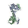
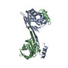
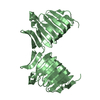
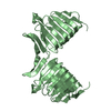
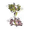
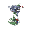
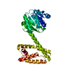
Archease / Archease domain / Archease domain superfamily / Archease protein family (MTH1598/TM1083) / tRNA processing / metal ion binding / DI(HYDROXYETHYL)ETHER / Protein archease
Function and homology information
Biological speciesHomo sapiens (human)
MethodX-RAY DIFFRACTION / SYNCHROTRON / MOLECULAR REPLACEMENT / Resolution: 1.97 Å
AuthorsDuan, S.Y. / Li, J.X.
Funding support China, 1items
OrganizationGrant numberCountry
China
CitationJournal: Int.J.Biochem.Cell Biol. / Year: 2020
Title: Crystal structure of human archease, a key cofactor of tRNA splicing ligase complex.
Authors: Duan, S. / Gao, W. / Chen, Z. / Li, Z. / Li, S. / Gan, J. / Chen, X. / Li, J.
History
DepositionDec 11, 2017Deposition site: PDBJ / Processing site: PDBJ
Revision 1.0Jan 2, 2019Provider: repository / Type: Initial release
Revision 1.1Jul 15, 2020Group: Database references / Category: citation / citation_author
Item: _citation.country / _citation.journal_abbrev ..._citation.country / _citation.journal_abbrev / _citation.journal_id_CSD / _citation.journal_id_ISSN / _citation.journal_volume / _citation.page_first / _citation.page_last / _citation.pdbx_database_id_DOI / _citation.pdbx_database_id_PubMed / _citation.title / _citation.year
Revision 1.2Nov 22, 2023Group: Data collection / Database references / Refinement description
Category: chem_comp_atom / chem_comp_bond ...chem_comp_atom / chem_comp_bond / database_2 / pdbx_initial_refinement_model
Item: _database_2.pdbx_DOI / _database_2.pdbx_database_accession

-
Structure visualization

Structure viewerMolecule:
MolmilJmol/JSmol

Downloads & links

-
Assembly

Deposited unit
A: Protein archease
B: Protein archease
hetero molecules


Theoretical massNumber of molelcules
Total (without water)43,9495
Polymers43,6752
Non-polymers2743
Water1,928107
1


  • Idetical with deposited unit
  • defined by author&software
  • Evidence: gel filtration, SEC-MALS for absolute molar mass and size measurement
TypeNameSymmetry operationNumber
identity operation1_555x,y,z1
Buried area5120 Å2
ΔGint-10 kcal/mol
Surface area16030 Å2
MethodPISA
Unit cell
Length a, b, c (Å)46.201, 55.844, 133.261
Angle α, β, γ (deg.)90.00, 90.00, 90.00
Int Tables number19
Space group name H-MP212121

-
Components

#1: Protein Protein archease / cDNA FLJ75987 / highly similar to Homo sapiens zinc finger and BTB domain containing 8 opposite ...cDNA FLJ75987 / highly similar to Homo sapiens zinc finger and BTB domain containing 8 opposite strand (ZBTB8OS) / mRNA


Mass: 21837.322 Da / Num. of mol.: 2
Source method: isolated from a genetically manipulated source
Source: (gene. exp.) Homo sapiens (human) / Gene: ZBTB8OS / Plasmid: pET28b(+) / Production host: Escherichia coli (E. coli) / References: UniProt: A8K0B5
#2: Chemical ChemComp-PEG / DI(HYDROXYETHYL)ETHER / Diethylene glycol


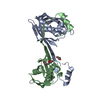
Mass: 106.120 Da / Num. of mol.: 2 / Source method: obtained synthetically / Formula: C4H10O3
#3: Chemical ChemComp-EDO / 1,2-ETHANEDIOL / ETHYLENE GLYCOL / Ethylene glycol


Mass: 62.068 Da / Num. of mol.: 1 / Source method: obtained synthetically / Formula: C2H6O2
#4: Water ChemComp-HOH / water / Water


Mass: 18.015 Da / Num. of mol.: 107 / Source method: isolated from a natural source / Formula: H2O

-
Experimental details

-
Experiment

ExperimentMethod: X-RAY DIFFRACTION / Number of used crystals: 1

-
Sample preparation

CrystalDensity Matthews: 1.97 Å3/Da / Density % sol: 37.5 %
Crystal growTemperature: 290 K / Method: vapor diffusion, hanging drop / pH: 5 / Details: 20% PEG 6000, 100mM sodium citrate pH5.0

-
Data collection

DiffractionMean temperature: 100 K
Diffraction sourceSource: SYNCHROTRON / Site: SSRF / Beamline: BL18U1 / Wavelength: 0.9778 Å
DetectorType: ADSC QUANTUM 315r / Detector: CCD / Date: Dec 19, 2015
RadiationProtocol: SINGLE WAVELENGTH / Monochromatic (M) / Laue (L): M / Scattering type: x-ray
Radiation wavelengthWavelength: 0.9778 Å / Relative weight: 1
ReflectionResolution: 1.961→50 Å / Num. obs: 24285 / % possible obs: 96.1 % / Redundancy: 11.5 % / Net I/σ(I): 19.778
Reflection shellResolution: 1.98→2.01 Å

-
Processing

Software
NameVersionClassification
REFMAC5.8.0135refinement
HKL-3000data reduction
HKL-3000data scaling
MLPHAREphasing
RefinementMethod to determine structure: MOLECULAR REPLACEMENT
Starting model: 4N2P
Resolution: 1.97→42.8 Å / Cross valid method: THROUGHOUT
RfactorNum. reflection% reflection
Rfree0.23395 1255 -
Rwork0.1839 --
obs-22975 96.6 %
Refinement stepCycle: LAST / Resolution: 1.97→42.8 Å
ProteinNucleic acidLigandSolventTotal
Num. atoms2625 0 18 107 2750

+
About Yorodumi

-
News

-
Feb 9, 2022. New format data for meta-information of EMDB entries

New format data for meta-information of EMDB entries

  • Version 3 of the EMDB header file is now the official format.
  • The previous official version 1.9 will be removed from the archive.

Related info.:EMDB header

External links:wwPDB to switch to version 3 of the EMDB data model

-
Aug 12, 2020. Covid-19 info

Covid-19 info

URL: https://pdbj.org/emnavi/covid19.php

New page: Covid-19 featured information page in EM Navigator.

Related info.:Covid-19 info / Mar 5, 2020. Novel coronavirus structure data

+
Mar 5, 2020. Novel coronavirus structure data

Novel coronavirus structure data

Related info.:Yorodumi Speices / Aug 12, 2020. Covid-19 info

External links:COVID-19 featured content - PDBj / Molecule of the Month (242):Coronavirus Proteases

+
Jan 31, 2019. EMDB accession codes are about to change! (news from PDBe EMDB page)

EMDB accession codes are about to change! (news from PDBe EMDB page)

  • The allocation of 4 digits for EMDB accession codes will soon come to an end. Whilst these codes will remain in use, new EMDB accession codes will include an additional digit and will expand incrementally as the available range of codes is exhausted. The current 4-digit format prefixed with “EMD-” (i.e. EMD-XXXX) will advance to a 5-digit format (i.e. EMD-XXXXX), and so on. It is currently estimated that the 4-digit codes will be depleted around Spring 2019, at which point the 5-digit format will come into force.
  • The EM Navigator/Yorodumi systems omit the EMD- prefix.

Related info.:Q: What is EMD? / ID/Accession-code notation in Yorodumi/EM Navigator

External links:EMDB Accession Codes are Changing Soon! / Contact to PDBj

+
Jul 12, 2017. Major update of PDB

Major update of PDB

  • wwPDB released updated PDB data conforming to the new PDBx/mmCIF dictionary.
  • This is a major update changing the version number from 4 to 5, and with Remediation, in which all the entries are updated.
  • In this update, many items about electron microscopy experimental information are reorganized (e.g. em_software).
  • Now, EM Navigator and Yorodumi are based on the updated data.

External links:wwPDB Remediation / Enriched Model Files Conforming to OneDep Data Standards Now Available in the PDB FTP Archive

-
Yorodumi

Thousand views of thousand structures

  • Yorodumi is a browser for structure data from EMDB, PDB, SASBDB, etc.
  • This page is also the successor to EM Navigator detail page, and also detail information page/front-end page for Omokage search.
  • The word "yorodu" (or yorozu) is an old Japanese word meaning "ten thousand". "mi" (miru) is to see.

Related info.:EMDB / PDB / SASBDB / Comparison of 3 databanks / Yorodumi Search / Aug 31, 2016. New EM Navigator & Yorodumi / Yorodumi Papers / Jmol/JSmol / Function and homology information / Changes in new EM Navigator and Yorodumi

Read more