 Movie
Movie Controller


+ Open data

- Basic information
| Entry | Database: PDB / ID: 1k4z | |||||||||
|---|---|---|---|---|---|---|---|---|---|---|
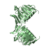
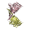
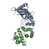
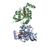
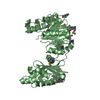
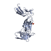
| Title | C-terminal Domain of Cyclase Associated Protein | |||||||||
 Components Components | Adenylyl Cyclase-Associated Protein | |||||||||
 Keywords Keywords | MEMBRANE PROTEIN / right-handed parallel beta-helix / intertwined dimer / actin-binding / New York SGX Research Center for Structural Genomics / NYSGXRC / Structural Genomics / PSI / Protein Structure Initiative | |||||||||
| Function / homology |  Function and homology information Function and homology informationpositive regulation of actin filament depolymerization / actin cortical patch / regulation of actin filament polymerization / actin filament severing / mating projection tip / actin filament depolymerization / positive regulation of Ras protein signal transduction / positive regulation of mitochondrial fission / adenylate cyclase binding / Neutrophil degranulation ...positive regulation of actin filament depolymerization / actin cortical patch / regulation of actin filament polymerization / actin filament severing / mating projection tip / actin filament depolymerization / positive regulation of Ras protein signal transduction / positive regulation of mitochondrial fission / adenylate cyclase binding / Neutrophil degranulation / actin filament organization / actin binding / Ras protein signal transduction / identical protein binding / cytoplasm Similarity search - Function | |||||||||
| Biological species |  | |||||||||
| Method |  X-RAY DIFFRACTION / SYNCHROTRON / MAD / Resolution: 2.3 Å X-RAY DIFFRACTION / SYNCHROTRON / MAD / Resolution: 2.3 Å | |||||||||
 Authors Authors | Rozwarski, D.A. / Fedorov, A.A. / Dodatko, T. / Almo, S.C. / Burley, S.K. / New York SGX Research Center for Structural Genomics (NYSGXRC) | |||||||||
 Citation Citation | Journal: Biochemistry / Year: 2004 Title: Crystal structure of the actin binding domain of the cyclase-associated protein Authors: Dodatko, T. / Fedorov, A.A. / Grynberg, M. / Patskovsky, Y. / Rozwarski, D.A. / Jaroszewski, L. / Aronoff-Spencer, E. / Kondraskina, E. / Irving, T. / Godzik, A. / Almo, S.C. #1: Journal: J.Biol.Chem. / Year: 1996Title: Two Separate Functions Are Encoded by the Carboxyl-terminal Domains of the Yeast Cyclase-associated Protein and Its Mammalian Homologs. Dimerization and Actin Binding. Authors: Zelicof, A. / Protopopov, V. / David, D. / Lin, X.Y. / Lusgarten, V. / Gerst, J.E. | |||||||||
| History |
|
- Structure visualization
| Structure viewer | Molecule: MolmilJmol/JSmol |
|---|
- Downloads & links
-Download
| PDBx/mmCIF format | 1k4z.cif.gz | 77.3 KB | Display | PDBx/mmCIF format |
|---|---|---|---|---|
| PDB format | pdb1k4z.ent.gz | 57.2 KB | Display | PDB format |
| PDBx/mmJSON format | 1k4z.json.gz | Tree view | PDBx/mmJSON format | |
| Others |  Other downloads Other downloads |
-Validation report
| Arichive directory | https://data.pdbj.org/pub/pdb/validation_reports/k4/1k4zftp://data.pdbj.org/pub/pdb/validation_reports/k4/1k4z | HTTPS FTP |
|---|
-Related structure data


-Links
 PDBj
PDBj


- Assembly
| Deposited unit | 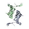
| ||||||||
|---|---|---|---|---|---|---|---|---|---|
| 1 | 
| ||||||||
| 2 | 
| ||||||||
| Unit cell |
| ||||||||
| Details | The biological assembly is an intertwined C-CAP dimer, which can be constructed using chain A and its symmetry partner generated by a crystallographic two-fold axis; or using chain B and its symmetry partner, generated by another crystallographic two-fold axis. |
-Components
| #1: Protein | Mass: 17540.641 Da / Num. of mol.: 2 / Fragment: C-TERMINAL DOMAIN Source method: isolated from a genetically manipulated source Source: (gene. exp.) Production host:  #2: Water | ChemComp-HOH / |  Mass: 18.015 Da / Num. of mol.: 160 / Source method: isolated from a natural source / Formula: H2O Mass: 18.015 Da / Num. of mol.: 160 / Source method: isolated from a natural source / Formula: H2OHas protein modification | Y | |
|---|
-Experimental details
-Experiment
| Experiment | Method: X-RAY DIFFRACTION / Number of used crystals: 1 |
|---|
- Sample preparation
| Crystal | Density Matthews: 2.827 Å3/Da / Density % sol: 56.53 % | ||||||||||||||||||||||||||||||
|---|---|---|---|---|---|---|---|---|---|---|---|---|---|---|---|---|---|---|---|---|---|---|---|---|---|---|---|---|---|---|---|
| Crystal grow | Temperature: 298 K / Method: vapor diffusion, hanging drop / pH: 7.6 Details: PEG 4000, lithium sulfate, HEPES, pH 7.6, VAPOR DIFFUSION, HANGING DROP, temperature 298.0K | ||||||||||||||||||||||||||||||
| Crystal grow | *PLUS Temperature: 16 ℃ / Method: vapor diffusion, sitting drop / pH: 8.5 | ||||||||||||||||||||||||||||||
| Components of the solutions | *PLUS
|
-Data collection
| Diffraction | Mean temperature: 103 K |
|---|---|
| Diffraction source | Source: SYNCHROTRON / Site: NSLS  / Beamline: X9B / Wavelength: 1.04 Å / Beamline: X9B / Wavelength: 1.04 Å |
| Detector | Type: MARRESEARCH / Detector: IMAGE PLATE / Date: Aug 30, 1998 |
| Radiation | Protocol: SINGLE WAVELENGTH / Monochromatic (M) / Laue (L): M / Scattering type: x-ray |
| Radiation wavelength | Wavelength: 1.04 Å / Relative weight: 1 |
| Reflection | Resolution: 2.3→10 Å / Num. all: 17249 / Num. obs: 17249 / % possible obs: 97.8 % / Observed criterion σ(F): 0 / Observed criterion σ(I): 0 / Redundancy: 6.1 % / Biso Wilson estimate: 37 Å2 / Rmerge(I) obs: 0.038 / Net I/σ(I): 49.7 |
| Reflection shell | Resolution: 2.3→2.44 Å / Redundancy: 5 % / Rmerge(I) obs: 0.106 / Mean I/σ(I) obs: 16.4 / Num. unique all: 2732 / % possible all: 92.9 |
| Reflection | *PLUS Highest resolution: 2.3 Å / Lowest resolution: 10 Å / Num. obs: 17479 / % possible obs: 97.3 % / Num. measured all: 105428 |
| Reflection shell | *PLUS Lowest resolution: 2.38 Å / % possible obs: 91.1 % / Num. unique obs: 1564 / Num. measured obs: 7805 / Rmerge(I) obs: 0.1 / Mean I/σ(I) obs: 18.1 |
- Processing
| Software |
| ||||||||||||||||||||||||||||||||||||
|---|---|---|---|---|---|---|---|---|---|---|---|---|---|---|---|---|---|---|---|---|---|---|---|---|---|---|---|---|---|---|---|---|---|---|---|---|---|
| Refinement | Method to determine structure: MAD / Resolution: 2.3→9.99 Å / Rfactor Rfree error: 0.006 / Cross valid method: THROUGHOUT / σ(F): 0 / Stereochemistry target values: Engh & Huber
| ||||||||||||||||||||||||||||||||||||
| Solvent computation | Solvent model: flat model / Bsol: 44.52 Å2 / ksol: 0.429148 e/Å3 | ||||||||||||||||||||||||||||||||||||
| Displacement parameters | Biso mean: 32.3 Å2 | ||||||||||||||||||||||||||||||||||||
| Refine analyze |
| ||||||||||||||||||||||||||||||||||||
| Refinement step | Cycle: LAST / Resolution: 2.3→9.99 Å
| ||||||||||||||||||||||||||||||||||||
| Refine LS restraints |
| ||||||||||||||||||||||||||||||||||||
| Refine LS restraints NCS | NCS model details: restraints | ||||||||||||||||||||||||||||||||||||
| LS refinement shell | Resolution: 2.3→2.44 Å / Rfactor Rfree error: 0.018 / Total num. of bins used: 6
| ||||||||||||||||||||||||||||||||||||
| Xplor file |
| ||||||||||||||||||||||||||||||||||||
| Refinement | *PLUS σ(F): 0 / % reflection Rfree: 10 % / Rfactor obs: 0.207 | ||||||||||||||||||||||||||||||||||||
| Solvent computation | *PLUS | ||||||||||||||||||||||||||||||||||||
| Displacement parameters | *PLUS Biso mean: 32.3 Å2 | ||||||||||||||||||||||||||||||||||||
| Refine LS restraints | *PLUS
| ||||||||||||||||||||||||||||||||||||
| LS refinement shell | *PLUS % reflection Rfree: 9.3 % / Rfactor Rwork: 0.224 / Rfactor obs: 0.224 |
