ISOMERASE / phosphoglycerate mutase / structural genomics / PSI-2 / protein genomics / MCSG / glycolysis isomerase / Protein Structure Initiative / Midwest Center for Structural Genomics / Glycolysis
Function / homology
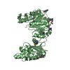
Function and homology information
phosphoglycerate mutase (2,3-diphosphoglycerate-independent) / phosphoglycerate mutase activity / glycolytic process / metal ion binding Similarity search - Function
Resolution: 2.6→73.92 Å / Num. obs: 25760 / % possible obs: 99.84 % / Observed criterion σ(F): 3 / Redundancy: 9.5 % / Rmerge(I) obs: 0.105 / Net I/σ(I): 30.75
Reflection shell
Resolution: 2.6→2.62 Å / Redundancy: 4.2 % / Rmerge(I) obs: 0.509 / Mean I/σ(I) obs: 2.923 / % possible all: 99.5
-
Processing
Software
Name
Version
Classification
SBC-Collect
datacollection
MLPHARE
phasing
REFMAC
5.5.0102
refinement
HKL-3000
datareduction
HKL-3000
datascaling
Refinement
Method to determine structure: MAD / Resolution: 2.6→73.92 Å / Cor.coef. Fo:Fc: 0.928 / Cor.coef. Fo:Fc free: 0.885 / SU B: 12.677 / SU ML: 0.276 / Cross valid method: THROUGHOUT / ESU R: 1.18 / ESU R Free: 0.371 / Stereochemistry target values: MAXIMUM LIKELIHOOD Details: HYDROGENS HAVE BEEN ADDED IN THE RIDING POSITIONS. Authors state that residue B58 and B270 do not have well-defined side chain because of weak electron density at the region.
Rfactor
Num. reflection
% reflection
Selection details
Rfree
0.28782
1305
5.1 %
RANDOM
Rwork
0.20737
-
-
-
obs
0.21145
24352
99.81 %
-
Solvent computation
Ion probe radii: 0.8 Å / Shrinkage radii: 0.8 Å / VDW probe radii: 1.4 Å / Solvent model: MASK
Displacement parameters
Biso mean: 33.771 Å2
Baniso -1
Baniso -2
Baniso -3
1-
0.16 Å2
0 Å2
0 Å2
2-
-
-2.3 Å2
0 Å2
3-
-
-
2.14 Å2
Refinement step
Cycle: LAST / Resolution: 2.6→73.92 Å
Protein
Nucleic acid
Ligand
Solvent
Total
Num. atoms
5616
0
0
145
5761
Refine LS restraints
Refine-ID
Type
Dev ideal
Dev ideal target
Number
X-RAY DIFFRACTION
r_bond_refined_d
0.014
0.022
5731
X-RAY DIFFRACTION
r_angle_refined_deg
1.643
1.982
7743
X-RAY DIFFRACTION
r_dihedral_angle_1_deg
7.276
5
723
X-RAY DIFFRACTION
r_dihedral_angle_2_deg
36.939
23.256
258
X-RAY DIFFRACTION
r_dihedral_angle_3_deg
20.488
15
982
X-RAY DIFFRACTION
r_dihedral_angle_4_deg
20.357
15
57
X-RAY DIFFRACTION
r_chiral_restr
0.114
0.2
853
X-RAY DIFFRACTION
r_gen_planes_refined
0.007
0.021
4369
X-RAY DIFFRACTION
r_mcbond_it
0.786
1.5
3633
X-RAY DIFFRACTION
r_mcangle_it
1.524
2
5836
X-RAY DIFFRACTION
r_scbond_it
2.447
3
2098
X-RAY DIFFRACTION
r_scangle_it
4.004
4.5
1907
LS refinement shell
Resolution: 2.598→2.665 Å / Total num. of bins used: 20
Rfactor
Num. reflection
% reflection
Rfree
0.356
111
-
Rwork
0.257
1758
-
obs
-
-
99.84 %
+
About Yorodumi
-
News
-
Feb 9, 2022. New format data for meta-information of EMDB entries
New format data for meta-information of EMDB entries
Version 3 of the EMDB header file is now the official format.
The previous official version 1.9 will be removed from the archive.
In the structure databanks used in Yorodumi, some data are registered as the other names, "COVID-19 virus" and "2019-nCoV". Here are the details of the virus and the list of structure data.
Jan 31, 2019. EMDB accession codes are about to change! (news from PDBe EMDB page)
EMDB accession codes are about to change! (news from PDBe EMDB page)
The allocation of 4 digits for EMDB accession codes will soon come to an end. Whilst these codes will remain in use, new EMDB accession codes will include an additional digit and will expand incrementally as the available range of codes is exhausted. The current 4-digit format prefixed with “EMD-” (i.e. EMD-XXXX) will advance to a 5-digit format (i.e. EMD-XXXXX), and so on. It is currently estimated that the 4-digit codes will be depleted around Spring 2019, at which point the 5-digit format will come into force.
The EM Navigator/Yorodumi systems omit the EMD- prefix.
Related info.:Q: What is EMD? / ID/Accession-code notation in Yorodumi/EM Navigator
Yorodumi is a browser for structure data from EMDB, PDB, SASBDB, etc.
This page is also the successor to EM Navigator detail page, and also detail information page/front-end page for Omokage search.
The word "yorodu" (or yorozu) is an old Japanese word meaning "ten thousand". "mi" (miru) is to see.
Related info.:EMDB / PDB / SASBDB / Comparison of 3 databanks / Yorodumi Search / Aug 31, 2016. New EM Navigator & Yorodumi / Yorodumi Papers / Jmol/JSmol / Function and homology information / Changes in new EM Navigator and Yorodumi
 Movie
Movie Controller

 Yorodumi
 Open data
 Basic information
 Components
 Keywords
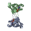
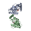
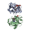
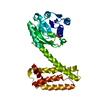
 Function and homology information
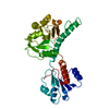
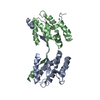
 Thermoplasma acidophilum (acidophilic)
 X-RAY DIFFRACTION /
  Authors
 Citation
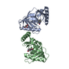
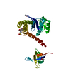
 Structure visualization
 Downloads & links
 Other downloads
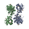

 PDBj
PDBj
 Assembly
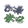

 Mass: 18.015 Da / Num. of mol.: 145 / Source method: isolated from a natural source / Formula: H2O
 Sample preparation
 / Beamline: 19-BM / Wavelength: 0.97880, 0.97904
 Processing