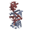
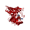
Entry Database : PDB / ID : 5a9rTitle Apo form of Imine reductase from Amycolatopsis orientalis IMINE REDUCTASE Keywords / / / Function / homology Function Domain/homology Component
/ / / / / / / / / / / / / / / / / / / / / / / / / / / / / / Biological species AMYCOLATOPSIS ORIENTALIS (bacteria)Method / / / Resolution : 1.55 Å Authors Man, H. / Aleku, G. / Turner, N.J. / Grogan, G. Journal : Acs Catalysis / Year : 2016Title : Stereoselectivity and Structural Characterization of an Imine Reductase (Ired) from Amycolatopsis OrientalisAuthors : Aleku, G.A. / Man, H. / France, S.P. / Leipold, F. / Hussain, S. / Toca-Gonzalez, L. / Marchington, R. / Hart, S. / Turkenburg, J.P. / Grogan, G. / Turner, N.J. History Deposition Jul 22, 2015 Deposition site / Processing site Revision 1.0 Jun 1, 2016 Provider / Type Revision 1.1 Jan 10, 2024 Group Data collection / Database references ... Data collection / Database references / Derived calculations / Other / Refinement description Category chem_comp_atom / chem_comp_bond ... chem_comp_atom / chem_comp_bond / database_2 / pdbx_database_status / pdbx_initial_refinement_model / struct_site Item _database_2.pdbx_DOI / _database_2.pdbx_database_accession ... _database_2.pdbx_DOI / _database_2.pdbx_database_accession / _pdbx_database_status.status_code_sf / _struct_site.pdbx_auth_asym_id / _struct_site.pdbx_auth_comp_id / _struct_site.pdbx_auth_seq_id
Show all Show less
 Movie
Movie Controller


 Open data
 Basic information
 Components
 Keywords
 Function and homology information
 AMYCOLATOPSIS ORIENTALIS (bacteria)
 X-RAY DIFFRACTION /
  Authors
 Citation
 Structure visualization
 Downloads & links
 Other downloads


 PDBj
PDBj
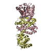
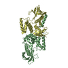
 Assembly


 Mass: 59.044 Da / Num. of mol.: 1 / Source method: obtained synthetically / Formula: C2H3O2
 Mass: 18.015 Da / Num. of mol.: 101 / Source method: isolated from a natural source / Formula: H2O
 Sample preparation
 / Beamline: I03 / Wavelength: 0.9795
  Processing