 Movie
Movie Controller

[English] 日本語
 Yorodumi
- PDB-3o8q: 1.45 Angstrom Resolution Crystal Structure of Shikimate 5-Dehydro... -
+ Open data

- Basic information
| Entry | Database: PDB / ID: 3o8q | |||||||||
|---|---|---|---|---|---|---|---|---|---|---|
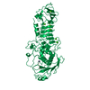
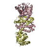
| Title | 1.45 Angstrom Resolution Crystal Structure of Shikimate 5-Dehydrogenase (aroE) from Vibrio cholerae | |||||||||
 Components Components | (Shikimate 5-dehydrogenase I ...) x 2 | |||||||||
 Keywords Keywords | OXIDOREDUCTASE / Structural Genomics / Center for Structural Genomics of Infectious Diseases / CSGID / alpha/beta domain | |||||||||
| Function / homology | Leucine Dehydrogenase, chain A, domain 1 / NAD(P)-binding Rossmann-like Domain / Rossmann fold / 3-Layer(aba) Sandwich / Alpha Beta / :  Function and homology information Function and homology information | |||||||||
| Biological species |  Vibrio cholerae biovar El Tor (bacteria)Vibrio cholerae O1 biovar El Tor (bacteria) Vibrio cholerae biovar El Tor (bacteria)Vibrio cholerae O1 biovar El Tor (bacteria) | |||||||||
| Method |  X-RAY DIFFRACTION / SYNCHROTRON / MOLECULAR REPLACEMENT / Resolution: 1.45 Å X-RAY DIFFRACTION / SYNCHROTRON / MOLECULAR REPLACEMENT / Resolution: 1.45 Å | |||||||||
 Authors Authors | Minasov, G. / Light, S.H. / Shuvalova, L. / Papazisi, L. / Anderson, W.F. / Center for Structural Genomics of Infectious Diseases (CSGID) | |||||||||
 Citation Citation | Journal: TO BE PUBLISHED Title: 1.45 Angstrom Resolution Crystal Structure of Shikimate 5-Dehydrogenase (aroE) from Vibrio cholerae. Authors: Minasov, G. / Light, S.H. / Shuvalova, L. / Papazisi, L. / Anderson, W.F. / Center for Structural Genomics of Infectious Diseases (CSGID) | |||||||||
| History |
|
- Structure visualization
| Structure viewer | Molecule: MolmilJmol/JSmol |
|---|
- Downloads & links
-Download
| PDBx/mmCIF format | 3o8q.cif.gz | 247.6 KB | Display | PDBx/mmCIF format |
|---|---|---|---|---|
| PDB format | pdb3o8q.ent.gz | 197.1 KB | Display | PDB format |
| PDBx/mmJSON format | 3o8q.json.gz | Tree view | PDBx/mmJSON format | |
| Others |  Other downloads Other downloads |
-Validation report
| Arichive directory | https://data.pdbj.org/pub/pdb/validation_reports/o8/3o8qftp://data.pdbj.org/pub/pdb/validation_reports/o8/3o8q | HTTPS FTP |
|---|
-Related structure data
| Related structure data |  1nytS S: Starting model for refinement |
|---|---|
| Similar structure data | |
| Other databases |


-Links
 PDBj
PDBj- Assembly
| Deposited unit | 
| ||||||||
|---|---|---|---|---|---|---|---|---|---|
| 1 |
| ||||||||
| Unit cell |
|
-Components
-Shikimate 5-dehydrogenase I ... , 2 types, 2 molecules AB
| #1: Protein | Mass: 30552.748 Da / Num. of mol.: 1 Source method: isolated from a genetically manipulated source Source: (gene. exp.) Vibrio cholerae biovar El Tor (bacteria)Strain: N16961 / Gene: aroE, VCD_001524 / Plasmid: pMCSG7 / Production host: References: UniProt: C3NTR0, shikimate dehydrogenase (NADP+) |
|---|---|
| #2: Protein | Mass: 29974.109 Da / Num. of mol.: 1 / Mutation: Q4(PCA) Source method: isolated from a genetically manipulated source Source: (gene. exp.) Vibrio cholerae O1 biovar El Tor (bacteria)Strain: N16961 / Gene: aroE, VCD_001524 / Plasmid: pMCSG7 / Production host: References: UniProt: C3NTR0, shikimate dehydrogenase (NADP+) |
-Non-polymers , 4 types, 528 molecules 


| #3: Chemical |  Mass: 22.990 Da / Num. of mol.: 2 / Source method: obtained synthetically / Formula: Na Mass: 22.990 Da / Num. of mol.: 2 / Source method: obtained synthetically / Formula: Na#4: Chemical | ChemComp-SO4 /  Mass: 96.063 Da / Num. of mol.: 4 / Source method: obtained synthetically / Formula: SO4 Mass: 96.063 Da / Num. of mol.: 4 / Source method: obtained synthetically / Formula: SO4#5: Chemical | ChemComp-EPE / |  Mass: 238.305 Da / Num. of mol.: 1 / Source method: obtained synthetically / Formula: C8H18N2O4S / Comment: pH buffer*YM Mass: 238.305 Da / Num. of mol.: 1 / Source method: obtained synthetically / Formula: C8H18N2O4S / Comment: pH buffer*YM#6: Water | ChemComp-HOH / | Mass: 18.015 Da / Num. of mol.: 521 / Source method: isolated from a natural source / Formula: H2O |
|---|
-Details
| Has protein modification | Y |
|---|
-Experimental details
-Experiment
| Experiment | Method: X-RAY DIFFRACTION / Number of used crystals: 1 |
|---|
- Sample preparation
| Crystal | Density Matthews: 2.03 Å3/Da / Density % sol: 39.4 % |
|---|---|
| Crystal grow | Temperature: 295 K / Method: vapor diffusion, sitting drop / pH: 7.5 Details: Protein: 6.8 mGr/mL, 0.5 Sodium cloride, 0.01M Tris-HCl pH 8.3, 10 mM Curcumin; Screen: PEGs II (D1), 0.1M Sodium acetate, 0.1M HEPES pH 7.5, 22% (w/v) PEG 4000, VAPOR DIFFUSION, SITTING DROP, temperature 295K |
-Data collection
| Diffraction | Mean temperature: 100 K |
|---|---|
| Diffraction source | Source: SYNCHROTRON / Site: APS  / Beamline: 21-ID-F / Wavelength: 0.97872 Å / Beamline: 21-ID-F / Wavelength: 0.97872 Å |
| Detector | Type: MARMOSAIC 225 mm CCD / Detector: CCD / Date: Jul 23, 2010 / Details: Beryllium lenses |
| Radiation | Monochromator: Diamond / Protocol: SINGLE WAVELENGTH / Monochromatic (M) / Laue (L): M / Scattering type: x-ray |
| Radiation wavelength | Wavelength: 0.97872 Å / Relative weight: 1 |
| Reflection | Resolution: 1.45→30 Å / Num. all: 88234 / Num. obs: 88234 / % possible obs: 99.8 % / Observed criterion σ(I): -3 / Redundancy: 5.8 % / Biso Wilson estimate: 16.4 Å2 / Rmerge(I) obs: 0.058 / Net I/σ(I): 25.7 |
| Reflection shell | Resolution: 1.45→1.48 Å / Redundancy: 5.6 % / Rmerge(I) obs: 0.549 / Mean I/σ(I) obs: 3.2 / Num. unique all: 4378 / % possible all: 100 |
- Processing
| Software |
| ||||||||||||||||||||||||||||||||||||||||||||||||||||||||||||||||||||||||||||||||||||||||||
|---|---|---|---|---|---|---|---|---|---|---|---|---|---|---|---|---|---|---|---|---|---|---|---|---|---|---|---|---|---|---|---|---|---|---|---|---|---|---|---|---|---|---|---|---|---|---|---|---|---|---|---|---|---|---|---|---|---|---|---|---|---|---|---|---|---|---|---|---|---|---|---|---|---|---|---|---|---|---|---|---|---|---|---|---|---|---|---|---|---|---|---|
| Refinement | Method to determine structure: MOLECULAR REPLACEMENT Starting model: 1NYT Resolution: 1.45→29.32 Å / Cor.coef. Fo:Fc: 0.969 / Cor.coef. Fo:Fc free: 0.954 / SU B: 2.25 / SU ML: 0.041 / Isotropic thermal model: Mixed, isotropic and anisotropic. / Cross valid method: THROUGHOUT / ESU R Free: 0.071 / Stereochemistry target values: MAXIMUM LIKELIHOOD / Details: HYDROGENS HAVE BEEN ADDED IN THE RIDING POSITIONS
| ||||||||||||||||||||||||||||||||||||||||||||||||||||||||||||||||||||||||||||||||||||||||||
| Solvent computation | Ion probe radii: 0.8 Å / Shrinkage radii: 0.8 Å / VDW probe radii: 1.2 Å / Solvent model: BABINET MODEL WITH MASK | ||||||||||||||||||||||||||||||||||||||||||||||||||||||||||||||||||||||||||||||||||||||||||
| Displacement parameters | Biso mean: 15.218 Å2
| ||||||||||||||||||||||||||||||||||||||||||||||||||||||||||||||||||||||||||||||||||||||||||
| Refinement step | Cycle: LAST / Resolution: 1.45→29.32 Å
| ||||||||||||||||||||||||||||||||||||||||||||||||||||||||||||||||||||||||||||||||||||||||||
| Refine LS restraints |
| ||||||||||||||||||||||||||||||||||||||||||||||||||||||||||||||||||||||||||||||||||||||||||
| LS refinement shell | Resolution: 1.45→1.488 Å / Total num. of bins used: 20
|
