 Movie
Movie Controller

[English] 日本語
 Yorodumi
- PDB-8brt: Crystal structure of a variant of penicillin G acylase from Bacil... -
+ Open data

- Basic information
| Entry | Database: PDB / ID: 8brt | ||||||
|---|---|---|---|---|---|---|---|
| Title | Crystal structure of a variant of penicillin G acylase from Bacillaceae i. s. sp. FJAT-27231 with reduced surface entropy and additionally engineered crystal contact | ||||||
 Components Components | (Penicillin G ...) x 2 | ||||||
 Keywords Keywords | HYDROLASE / Penicillin / Acylase / Surface entropy reduction / Crystal contact engineering / Crystallizability | ||||||
| Function / homology |  Function and homology information Function and homology informationhydrolase activity, acting on carbon-nitrogen (but not peptide) bonds, in linear amides / antibiotic biosynthetic process / metal ion binding Similarity search - Function | ||||||
| Biological species |  | ||||||
| Method |  X-RAY DIFFRACTION / SYNCHROTRON / MOLECULAR REPLACEMENT / Resolution: 1.31 Å X-RAY DIFFRACTION / SYNCHROTRON / MOLECULAR REPLACEMENT / Resolution: 1.31 Å | ||||||
 Authors Authors | Wichmann, J. / Mayer, J. / Mattes, H. / Lukat, P. / Blankenfeldt, W. / Biedendieck, R. | ||||||
| Funding support |  Germany, 1items Germany, 1items
| ||||||
 Citation Citation | Journal: Cryst.Growth Des. / Year: 2023 Title: Multistep Engineering of a Penicillin G Acylase for Systematic Improvement of Crystallization Efficiency Authors: Wichmann, J. / Mayer, J. / Hintmann, M. / Lukat, P. / Blankenfeldt, W. / Biedendieck, R. | ||||||
| History |
|
- Structure visualization
| Structure viewer | Molecule: MolmilJmol/JSmol |
|---|
- Downloads & links
-Download
| PDBx/mmCIF format | 8brt.cif.gz | 442.7 KB | Display | PDBx/mmCIF format |
|---|---|---|---|---|
| PDB format | pdb8brt.ent.gz | 364.6 KB | Display | PDB format |
| PDBx/mmJSON format | 8brt.json.gz | Tree view | PDBx/mmJSON format | |
| Others |  Other downloads Other downloads |
-Validation report
| Arichive directory | https://data.pdbj.org/pub/pdb/validation_reports/br/8brtftp://data.pdbj.org/pub/pdb/validation_reports/br/8brt | HTTPS FTP |
|---|
-Related structure data


-Links
 PDBj
PDBj


- Assembly
| Deposited unit | 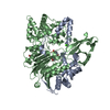
| ||||||||
|---|---|---|---|---|---|---|---|---|---|
| 1 |
| ||||||||
| Unit cell |
|
-Components
-Penicillin G ... , 2 types, 2 molecules AB
| #1: Protein | Mass: 24681.822 Da / Num. of mol.: 1 / Mutation: T167V, K201A, K202A, E203A Source method: isolated from a genetically manipulated source Details: penicillin G acylase, alpha-subunit, Surface entropy reduction variant (variant hypho_7) Source: (gene. exp.) Priestia megaterium (bacteria) / References: UniProt: A0A0K9H482 |
|---|---|
| #2: Protein | Mass: 61416.914 Da / Num. of mol.: 1 / Mutation: G14A, K135A, E136A, K137A, K403A, E404A, E405A Source method: isolated from a genetically manipulated source Details: penicillin G acylase, beta-subunit, Surface entropy reduction variant (variant hypho_7) Source: (gene. exp.) Priestia megaterium (bacteria) / References: UniProt: A0A0K9H482 |
-Non-polymers , 4 types, 822 molecules 


| #3: Chemical | ChemComp-EPE /  Mass: 238.305 Da / Num. of mol.: 1 / Source method: obtained synthetically / Formula: C8H18N2O4S / Comment: pH buffer*YM Mass: 238.305 Da / Num. of mol.: 1 / Source method: obtained synthetically / Formula: C8H18N2O4S / Comment: pH buffer*YM | ||||
|---|---|---|---|---|---|
| #4: Chemical |  Mass: 90.121 Da / Num. of mol.: 2 / Source method: obtained synthetically / Formula: C4H10O2 Mass: 90.121 Da / Num. of mol.: 2 / Source method: obtained synthetically / Formula: C4H10O2#5: Chemical |  Mass: 40.078 Da / Num. of mol.: 2 / Source method: obtained synthetically / Formula: Ca Mass: 40.078 Da / Num. of mol.: 2 / Source method: obtained synthetically / Formula: Ca#6: Water | ChemComp-HOH / | Mass: 18.015 Da / Num. of mol.: 817 / Source method: isolated from a natural source / Formula: H2O |
-Details
| Has ligand of interest | N |
|---|
-Experimental details
-Experiment
| Experiment | Method: X-RAY DIFFRACTION / Number of used crystals: 1 |
|---|
- Sample preparation
| Crystal | Density Matthews: 2.51 Å3/Da / Density % sol: 51.07 % |
|---|---|
| Crystal grow | Temperature: 293 K / Method: microbatch / pH: 7.5 Details: 100 mM HEPES pH 7.5 PEG 8000 Microbatch under paraffin oil |
-Data collection
| Diffraction | Mean temperature: 100 K / Serial crystal experiment: N |
|---|---|
| Diffraction source | Source: SYNCHROTRON / Site: PETRA III, DESY / Beamline: P11 / Wavelength: 1.0332 Å |
| Detector | Type: DECTRIS EIGER X 16M / Detector: PIXEL / Date: Sep 8, 2021 |
| Radiation | Protocol: SINGLE WAVELENGTH / Monochromatic (M) / Laue (L): M / Scattering type: x-ray |
| Radiation wavelength | Wavelength: 1.0332 Å / Relative weight: 1 |
| Reflection | Resolution: 1.31→81.92 Å / Num. obs: 208981 / % possible obs: 99.8 % / Redundancy: 10 % / CC1/2: 0.999 / Rmerge(I) obs: 0.05 / Rpim(I) all: 0.017 / Rrim(I) all: 0.052 / Χ2: 0.84 / Net I/σ(I): 19.8 / Num. measured all: 2081190 |
| Reflection shell | Resolution: 1.31→1.38 Å / % possible obs: 99.9 % / Redundancy: 10 % / Rmerge(I) obs: 0.713 / Num. measured all: 303662 / Num. unique obs: 30239 / CC1/2: 0.859 / Rpim(I) all: 0.233 / Rrim(I) all: 0.751 / Χ2: 0.56 / Net I/σ(I) obs: 2.8 |
- Processing
| Software |
| |||||||||||||||||||||||||||||||||||||||||||||||||||||||||||||||||||||||||||||||||||||||||||||||||||||||||||||||||||||||||||||||||||||||||||||||||||||||||||||||||||||||||||||||||||||||||||||||||||||||||||||||||||||||||||||||||||||||||||||||||||||||||||||||||||||||||||||||||||
|---|---|---|---|---|---|---|---|---|---|---|---|---|---|---|---|---|---|---|---|---|---|---|---|---|---|---|---|---|---|---|---|---|---|---|---|---|---|---|---|---|---|---|---|---|---|---|---|---|---|---|---|---|---|---|---|---|---|---|---|---|---|---|---|---|---|---|---|---|---|---|---|---|---|---|---|---|---|---|---|---|---|---|---|---|---|---|---|---|---|---|---|---|---|---|---|---|---|---|---|---|---|---|---|---|---|---|---|---|---|---|---|---|---|---|---|---|---|---|---|---|---|---|---|---|---|---|---|---|---|---|---|---|---|---|---|---|---|---|---|---|---|---|---|---|---|---|---|---|---|---|---|---|---|---|---|---|---|---|---|---|---|---|---|---|---|---|---|---|---|---|---|---|---|---|---|---|---|---|---|---|---|---|---|---|---|---|---|---|---|---|---|---|---|---|---|---|---|---|---|---|---|---|---|---|---|---|---|---|---|---|---|---|---|---|---|---|---|---|---|---|---|---|---|---|---|---|---|---|---|---|---|---|---|---|---|---|---|---|---|---|---|---|---|---|---|---|---|---|---|---|---|---|---|---|---|---|---|---|---|---|---|---|---|---|---|---|---|---|---|---|---|---|---|---|---|---|
| Refinement | Method to determine structure: MOLECULAR REPLACEMENT / Resolution: 1.31→46.05 Å / SU ML: 0.11 / Cross valid method: FREE R-VALUE / σ(F): 1.36 / Phase error: 15.23 / Stereochemistry target values: ML
| |||||||||||||||||||||||||||||||||||||||||||||||||||||||||||||||||||||||||||||||||||||||||||||||||||||||||||||||||||||||||||||||||||||||||||||||||||||||||||||||||||||||||||||||||||||||||||||||||||||||||||||||||||||||||||||||||||||||||||||||||||||||||||||||||||||||||||||||||||
| Solvent computation | Shrinkage radii: 0.9 Å / VDW probe radii: 1.1 Å / Solvent model: FLAT BULK SOLVENT MODEL | |||||||||||||||||||||||||||||||||||||||||||||||||||||||||||||||||||||||||||||||||||||||||||||||||||||||||||||||||||||||||||||||||||||||||||||||||||||||||||||||||||||||||||||||||||||||||||||||||||||||||||||||||||||||||||||||||||||||||||||||||||||||||||||||||||||||||||||||||||
| Refinement step | Cycle: LAST / Resolution: 1.31→46.05 Å
| |||||||||||||||||||||||||||||||||||||||||||||||||||||||||||||||||||||||||||||||||||||||||||||||||||||||||||||||||||||||||||||||||||||||||||||||||||||||||||||||||||||||||||||||||||||||||||||||||||||||||||||||||||||||||||||||||||||||||||||||||||||||||||||||||||||||||||||||||||
| Refine LS restraints |
| |||||||||||||||||||||||||||||||||||||||||||||||||||||||||||||||||||||||||||||||||||||||||||||||||||||||||||||||||||||||||||||||||||||||||||||||||||||||||||||||||||||||||||||||||||||||||||||||||||||||||||||||||||||||||||||||||||||||||||||||||||||||||||||||||||||||||||||||||||
| LS refinement shell |
| |||||||||||||||||||||||||||||||||||||||||||||||||||||||||||||||||||||||||||||||||||||||||||||||||||||||||||||||||||||||||||||||||||||||||||||||||||||||||||||||||||||||||||||||||||||||||||||||||||||||||||||||||||||||||||||||||||||||||||||||||||||||||||||||||||||||||||||||||||
| Refinement TLS params. | Method: refined / Refine-ID: X-RAY DIFFRACTION
| |||||||||||||||||||||||||||||||||||||||||||||||||||||||||||||||||||||||||||||||||||||||||||||||||||||||||||||||||||||||||||||||||||||||||||||||||||||||||||||||||||||||||||||||||||||||||||||||||||||||||||||||||||||||||||||||||||||||||||||||||||||||||||||||||||||||||||||||||||
| Refinement TLS group |
|
