 Movie
Movie Controller

[English] 日本語
 Yorodumi
- PDB-8bjj: ExoY Nucleotidyl Cyclase domain from Vibrio nigripulchritudo MART... -
+ Open data

- Basic information
| Entry | Database: PDB / ID: 8bjj | ||||||
|---|---|---|---|---|---|---|---|
| Title | ExoY Nucleotidyl Cyclase domain from Vibrio nigripulchritudo MARTX toxin, bound to ATP-Mg-actin, human profilin 1 and a sulfate ion | ||||||
 Components Components |
| ||||||
 Keywords Keywords | TOXIN / bacterial nucleotidyl cyclase toxin / activated complex | ||||||
| Function / homology |  Function and homology information Function and homology informationcalcium- and calmodulin-responsive adenylate cyclase activity / synapse maturation / adenyl-nucleotide exchange factor activity / negative regulation of actin filament bundle assembly / modification of postsynaptic actin cytoskeleton / positive regulation of actin filament bundle assembly / adenylate cyclase / regulation of actin filament polymerization / Signaling by ROBO receptors / negative regulation of stress fiber assembly ...calcium- and calmodulin-responsive adenylate cyclase activity / synapse maturation / adenyl-nucleotide exchange factor activity / negative regulation of actin filament bundle assembly / modification of postsynaptic actin cytoskeleton / positive regulation of actin filament bundle assembly / adenylate cyclase / regulation of actin filament polymerization / Signaling by ROBO receptors / negative regulation of stress fiber assembly / proline-rich region binding / PCP/CE pathway / cytoskeletal motor activator activity / host cell cytosol / positive regulation of ruffle assembly / myosin heavy chain binding / tropomyosin binding / actin filament bundle / troponin I binding / filamentous actin / mesenchyme migration / positive regulation of actin filament polymerization / skeletal muscle myofibril / positive regulation of epithelial cell migration / actin filament bundle assembly / striated muscle thin filament / skeletal muscle thin filament assembly / actin monomer binding / skeletal muscle fiber development / stress fiber / phosphotyrosine residue binding / titin binding / actin filament polymerization / phosphatidylinositol-4,5-bisphosphate binding / filopodium / actin filament / neural tube closure / RHO GTPases Activate Formins / modulation of chemical synaptic transmission / Hydrolases; Acting on acid anhydrides; Acting on acid anhydrides to facilitate cellular and subcellular movement / small GTPase binding / calcium-dependent protein binding / Platelet degranulation / lamellipodium / toxin activity / transferase activity / actin binding / actin cytoskeleton organization / cell body / blood microparticle / cell cortex / cytoskeleton / protein stabilization / cadherin binding / protein domain specific binding / cysteine-type endopeptidase activity / focal adhesion / hydrolase activity / calcium ion binding / positive regulation of gene expression / regulation of transcription by RNA polymerase II / host cell plasma membrane / glutamatergic synapse / magnesium ion binding / proteolysis / RNA binding / extracellular exosome / extracellular region / ATP binding / membrane / metal ion binding / identical protein binding / nucleus / cytoplasm / cytosol Similarity search - Function | ||||||
| Biological species |  Homo sapiens (human) Homo sapiens (human) Vibrio nigripulchritudo (bacteria) Vibrio nigripulchritudo (bacteria) | ||||||
| Method |  X-RAY DIFFRACTION / SYNCHROTRON / MOLECULAR REPLACEMENT / Resolution: 1.699 Å X-RAY DIFFRACTION / SYNCHROTRON / MOLECULAR REPLACEMENT / Resolution: 1.699 Å | ||||||
 Authors Authors | Teixeira-Nunes, M. / Renault, L. / Retailleau, P. | ||||||
| Funding support |  France, 1items France, 1items
| ||||||
 Citation Citation | Journal: Plos Pathog. / Year: 2023 Title: Functional and structural insights into the multi-step activation and catalytic mechanism of bacterial ExoY nucleotidyl cyclase toxins bound to actin-profilin. Authors: Teixeira-Nunes, M. / Retailleau, P. / Raoux-Barbot, D. / Comisso, M. / Missinou, A.A. / Velours, C. / Plancqueel, S. / Ladant, D. / Mechold, U. / Renault, L. | ||||||
| History |
|
- Structure visualization
| Structure viewer | Molecule: MolmilJmol/JSmol |
|---|
- Downloads & links
-Download
| PDBx/mmCIF format | 8bjj.cif.gz | 375.4 KB | Display | PDBx/mmCIF format |
|---|---|---|---|---|
| PDB format | pdb8bjj.ent.gz | 299.9 KB | Display | PDB format |
| PDBx/mmJSON format | 8bjj.json.gz | Tree view | PDBx/mmJSON format | |
| Others |  Other downloads Other downloads |
-Validation report
| Arichive directory | https://data.pdbj.org/pub/pdb/validation_reports/bj/8bjjftp://data.pdbj.org/pub/pdb/validation_reports/bj/8bjj | HTTPS FTP |
|---|
-Related structure data
| Related structure data |  8bjhC  8bjiC 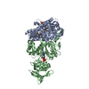 8bo1C  8br0C  8br1C  2pavS  7pj0 7pk7 7pqj S: Starting model for refinement C: citing same article ( |
|---|---|
| Similar structure data |
-Links
 PDBj
PDBj


- Assembly
| Deposited unit | 
| ||||||||
|---|---|---|---|---|---|---|---|---|---|
| 1 |
| ||||||||
| Unit cell |
|
-Components
-Protein , 3 types, 3 molecules ACB
| #1: Protein | Mass: 41862.613 Da / Num. of mol.: 1 / Source method: isolated from a natural source / Source: (natural) |
|---|---|
| #2: Protein | Mass: 14940.021 Da / Num. of mol.: 1 Source method: isolated from a genetically manipulated source Source: (gene. exp.) Homo sapiens (human) / Gene: PFN1 / Production host: |
| #3: Protein | Mass: 46247.477 Da / Num. of mol.: 1 Source method: isolated from a genetically manipulated source Source: (gene. exp.) Vibrio nigripulchritudo (bacteria) / Cell (production host): BL21(DE3) / Production host: |
-Non-polymers , 9 types, 735 molecules 


| #4: Chemical | ChemComp-ATP /  Mass: 507.181 Da / Num. of mol.: 1 / Source method: obtained synthetically / Formula: C10H16N5O13P3 / Feature type: SUBJECT OF INVESTIGATION / Comment: ATP, energy-carrying molecule*YM Mass: 507.181 Da / Num. of mol.: 1 / Source method: obtained synthetically / Formula: C10H16N5O13P3 / Feature type: SUBJECT OF INVESTIGATION / Comment: ATP, energy-carrying molecule*YM | ||||||||||
|---|---|---|---|---|---|---|---|---|---|---|---|
| #5: Chemical | ChemComp-MG /  Mass: 24.305 Da / Num. of mol.: 1 / Source method: obtained synthetically / Formula: Mg / Feature type: SUBJECT OF INVESTIGATION Mass: 24.305 Da / Num. of mol.: 1 / Source method: obtained synthetically / Formula: Mg / Feature type: SUBJECT OF INVESTIGATION | ||||||||||
| #6: Chemical | ChemComp-LAB /  Mass: 395.513 Da / Num. of mol.: 1 / Source method: obtained synthetically / Formula: C20H29NO5S / Feature type: SUBJECT OF INVESTIGATION Mass: 395.513 Da / Num. of mol.: 1 / Source method: obtained synthetically / Formula: C20H29NO5S / Feature type: SUBJECT OF INVESTIGATION | ||||||||||
| #7: Chemical |  Mass: 122.143 Da / Num. of mol.: 2 / Source method: obtained synthetically / Formula: C4H12NO3 / Comment: pH buffer*YM Mass: 122.143 Da / Num. of mol.: 2 / Source method: obtained synthetically / Formula: C4H12NO3 / Comment: pH buffer*YM#8: Chemical | ChemComp-SO4 /  Mass: 96.063 Da / Num. of mol.: 4 / Source method: obtained synthetically / Formula: SO4 / Feature type: SUBJECT OF INVESTIGATION Mass: 96.063 Da / Num. of mol.: 4 / Source method: obtained synthetically / Formula: SO4 / Feature type: SUBJECT OF INVESTIGATION#9: Chemical |  Mass: 34.015 Da / Num. of mol.: 2 / Source method: obtained synthetically / Formula: H2O2 Mass: 34.015 Da / Num. of mol.: 2 / Source method: obtained synthetically / Formula: H2O2#10: Chemical | ChemComp-PG4 / |  Mass: 194.226 Da / Num. of mol.: 1 / Source method: obtained synthetically / Formula: C8H18O5 / Comment: precipitant*YM Mass: 194.226 Da / Num. of mol.: 1 / Source method: obtained synthetically / Formula: C8H18O5 / Comment: precipitant*YM#11: Chemical | ChemComp-GOL / |  Mass: 92.094 Da / Num. of mol.: 1 / Source method: obtained synthetically / Formula: C3H8O3 Mass: 92.094 Da / Num. of mol.: 1 / Source method: obtained synthetically / Formula: C3H8O3#12: Water | ChemComp-HOH / | Mass: 18.015 Da / Num. of mol.: 722 / Source method: isolated from a natural source / Formula: H2O |
-Details
| Has ligand of interest | Y |
|---|
-Experimental details
-Experiment
| Experiment | Method: X-RAY DIFFRACTION / Number of used crystals: 1 |
|---|
- Sample preparation
| Crystal | Density Matthews: 2.36 Å3/Da / Density % sol: 47.88 % |
|---|---|
| Crystal grow | Temperature: 291 K / Method: vapor diffusion, sitting drop / pH: 8.5 / Details: 30% peg3000 0.3M LiSO4 0.1M Tris pH8.5 3% Dioxane |
-Data collection
| Diffraction | Mean temperature: 100 K / Serial crystal experiment: N |
|---|---|
| Diffraction source | Source: SYNCHROTRON / Site: SOLEIL / Beamline: PROXIMA 2 / Wavelength: 0.98 Å |
| Detector | Type: DECTRIS EIGER X 9M / Detector: PIXEL / Date: Dec 6, 2020 |
| Radiation | Protocol: SINGLE WAVELENGTH / Monochromatic (M) / Laue (L): M / Scattering type: x-ray |
| Radiation wavelength | Wavelength: 0.98 Å / Relative weight: 1 |
| Reflection | Resolution: 1.699→44.2 Å / Num. obs: 104207 / % possible obs: 99.8 % / Redundancy: 15.2 % / Biso Wilson estimate: 27.04 Å2 / Rmerge(I) obs: 0.057 / Net I/σ(I): 25.3 |
| Reflection shell | Resolution: 1.7→1.73 Å / Rmerge(I) obs: 1.055 / Mean I/σ(I) obs: 2.7 / Num. unique obs: 5211 |
- Processing
| Software |
| ||||||||||||||||||||||||||||||||||||||||||||||||||||||||||||||||||||||||||||||||||||||||||||||||||||||||||||
|---|---|---|---|---|---|---|---|---|---|---|---|---|---|---|---|---|---|---|---|---|---|---|---|---|---|---|---|---|---|---|---|---|---|---|---|---|---|---|---|---|---|---|---|---|---|---|---|---|---|---|---|---|---|---|---|---|---|---|---|---|---|---|---|---|---|---|---|---|---|---|---|---|---|---|---|---|---|---|---|---|---|---|---|---|---|---|---|---|---|---|---|---|---|---|---|---|---|---|---|---|---|---|---|---|---|---|---|---|---|
| Refinement | Method to determine structure: MOLECULAR REPLACEMENT Starting model: 2PAV Resolution: 1.699→44.2 Å / Cor.coef. Fo:Fc: 0.962 / Cor.coef. Fo:Fc free: 0.954 / SU R Cruickshank DPI: 0.097 / Cross valid method: THROUGHOUT / σ(F): 0 / SU R Blow DPI: 0.1 / SU Rfree Blow DPI: 0.092 / SU Rfree Cruickshank DPI: 0.09
| ||||||||||||||||||||||||||||||||||||||||||||||||||||||||||||||||||||||||||||||||||||||||||||||||||||||||||||
| Displacement parameters | Biso max: 110.7 Å2 / Biso mean: 33.36 Å2 / Biso min: 11.21 Å2
| ||||||||||||||||||||||||||||||||||||||||||||||||||||||||||||||||||||||||||||||||||||||||||||||||||||||||||||
| Refine analyze | Luzzati coordinate error obs: 0.2 Å | ||||||||||||||||||||||||||||||||||||||||||||||||||||||||||||||||||||||||||||||||||||||||||||||||||||||||||||
| Refinement step | Cycle: final / Resolution: 1.699→44.2 Å
| ||||||||||||||||||||||||||||||||||||||||||||||||||||||||||||||||||||||||||||||||||||||||||||||||||||||||||||
| Refine LS restraints |
| ||||||||||||||||||||||||||||||||||||||||||||||||||||||||||||||||||||||||||||||||||||||||||||||||||||||||||||
| LS refinement shell | Resolution: 1.7→1.71 Å / Rfactor Rfree error: 0 / Total num. of bins used: 51
| ||||||||||||||||||||||||||||||||||||||||||||||||||||||||||||||||||||||||||||||||||||||||||||||||||||||||||||
| Refinement TLS params. | Method: refined / Refine-ID: X-RAY DIFFRACTION
| ||||||||||||||||||||||||||||||||||||||||||||||||||||||||||||||||||||||||||||||||||||||||||||||||||||||||||||
| Refinement TLS group |
|
