 Movie
Movie Controller


+ Open data

- Basic information
| Entry | Database: PDB / ID: 8bib | ||||||
|---|---|---|---|---|---|---|---|
| Title | O-Methyltransferase Plu4890 in complex with SAH and AQ-256 | ||||||
 Components Components | Methyltransferase Plu4890 | ||||||
 Keywords Keywords | TRANSFERASE / methyltransferase / polyketide / anthraquinone | ||||||
| Function / homology |  Function and homology information Function and homology information | ||||||
| Biological species |  Photorhabdus laumondii subsp. laumondii TTO1 (bacteria) Photorhabdus laumondii subsp. laumondii TTO1 (bacteria) | ||||||
| Method |  X-RAY DIFFRACTION / SYNCHROTRON / MOLECULAR REPLACEMENT / Resolution: 2.3 Å X-RAY DIFFRACTION / SYNCHROTRON / MOLECULAR REPLACEMENT / Resolution: 2.3 Å | ||||||
 Authors Authors | Huber, E.M. / Groll, M. | ||||||
| Funding support | 1items
| ||||||
 Citation Citation | Journal: Structure / Year: 2023 Title: A set of closely related methyltransferases for site-specific tailoring of anthraquinone pigments. Authors: Huber, E.M. / Kreling, L. / Heinrich, A.K. / Dunnebacke, M. / Pothig, A. / Bode, H.B. / Groll, M. | ||||||
| History |
|
- Structure visualization
| Structure viewer | Molecule: MolmilJmol/JSmol |
|---|
- Downloads & links
-Download
| PDBx/mmCIF format | 8bib.cif.gz | 546.1 KB | Display | PDBx/mmCIF format |
|---|---|---|---|---|
| PDB format | pdb8bib.ent.gz | 453.6 KB | Display | PDB format |
| PDBx/mmJSON format | 8bib.json.gz | Tree view | PDBx/mmJSON format | |
| Others |  Other downloads Other downloads |
-Validation report
| Summary document | 8bib_validation.pdf.gz | 2.4 MB | Display | wwPDB validaton report |
|---|---|---|---|---|
| Full document | 8bib_full_validation.pdf.gz | 2.2 MB | Display | |
| Data in XML | 8bib_validation.xml.gz | 49.1 KB | Display | |
| Data in CIF | 8bib_validation.cif.gz | 68.4 KB | Display | |
| Arichive directory | https://data.pdbj.org/pub/pdb/validation_reports/bi/8bibftp://data.pdbj.org/pub/pdb/validation_reports/bi/8bib | HTTPS FTP |
-Related structure data
| Related structure data |  8bgtSC 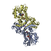 8bgxC 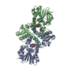 8bgyC 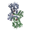 8bgzC 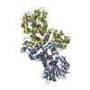 8bh0C 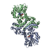 8bicC 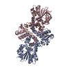 8bidC 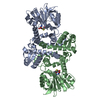 8bieC 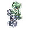 8bifC 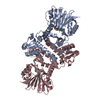 8bigC 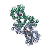 8bihC 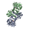 8biiC 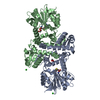 8bijC 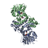 8birC S: Starting model for refinement C: citing same article ( |
|---|---|
| Similar structure data |
-Links
 PDBj
PDBj


- Assembly
| Deposited unit | 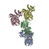
| ||||||||
|---|---|---|---|---|---|---|---|---|---|
| 1 | 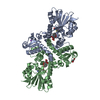
| ||||||||
| 2 | 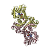
| ||||||||
| Unit cell |
|
-Components
-Protein , 1 types, 4 molecules ABCD
| #1: Protein | Mass: 36550.527 Da / Num. of mol.: 4 Source method: isolated from a genetically manipulated source Source: (gene. exp.) Photorhabdus laumondii subsp. laumondii TTO1 (bacteria)Production host: |
|---|
-Non-polymers , 7 types, 507 molecules 
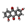


| #2: Chemical | ChemComp-SAH /  Type: L-peptide linking / Mass: 384.411 Da / Num. of mol.: 4 / Source method: obtained synthetically / Formula: C14H20N6O5S Type: L-peptide linking / Mass: 384.411 Da / Num. of mol.: 4 / Source method: obtained synthetically / Formula: C14H20N6O5S#3: Chemical | ChemComp-QOI /  Mass: 256.210 Da / Num. of mol.: 4 / Source method: obtained synthetically / Formula: C14H8O5 / Feature type: SUBJECT OF INVESTIGATION Mass: 256.210 Da / Num. of mol.: 4 / Source method: obtained synthetically / Formula: C14H8O5 / Feature type: SUBJECT OF INVESTIGATION#4: Chemical | ChemComp-NA / |  Mass: 22.990 Da / Num. of mol.: 1 / Source method: obtained synthetically / Formula: Na Mass: 22.990 Da / Num. of mol.: 1 / Source method: obtained synthetically / Formula: Na#5: Chemical | ChemComp-CO3 /  Mass: 60.009 Da / Num. of mol.: 4 / Source method: obtained synthetically / Formula: CO3 Mass: 60.009 Da / Num. of mol.: 4 / Source method: obtained synthetically / Formula: CO3#6: Chemical | ChemComp-GOL / |  Mass: 92.094 Da / Num. of mol.: 1 / Source method: obtained synthetically / Formula: C3H8O3 Mass: 92.094 Da / Num. of mol.: 1 / Source method: obtained synthetically / Formula: C3H8O3#7: Chemical |  Mass: 35.453 Da / Num. of mol.: 3 / Source method: obtained synthetically / Formula: Cl Mass: 35.453 Da / Num. of mol.: 3 / Source method: obtained synthetically / Formula: Cl#8: Water | ChemComp-HOH / | Mass: 18.015 Da / Num. of mol.: 490 / Source method: isolated from a natural source / Formula: H2O |
|---|
-Details
| Has ligand of interest | Y |
|---|
-Experimental details
-Experiment
| Experiment | Method: X-RAY DIFFRACTION / Number of used crystals: 1 |
|---|
- Sample preparation
| Crystal | Density Matthews: 2.77 Å3/Da / Density % sol: 55.53 % |
|---|---|
| Crystal grow | Temperature: 293 K / Method: vapor diffusion, sitting drop / pH: 6.5 Details: 0.2 M NaI, 0.1 M bis-tris propane pH 6.5, 20% PEG3350 |
-Data collection
| Diffraction | Mean temperature: 100 K / Serial crystal experiment: N |
|---|---|
| Diffraction source | Source: SYNCHROTRON / Site: SLS  / Beamline: X06SA / Wavelength: 1 Å / Beamline: X06SA / Wavelength: 1 Å |
| Detector | Type: DECTRIS EIGER X 16M / Detector: PIXEL / Date: Apr 14, 2018 |
| Radiation | Protocol: SINGLE WAVELENGTH / Monochromatic (M) / Laue (L): M / Scattering type: x-ray |
| Radiation wavelength | Wavelength: 1 Å / Relative weight: 1 |
| Reflection | Resolution: 2.3→48 Å / Num. obs: 68830 / % possible obs: 96.7 % / Redundancy: 3.1 % / Rmerge(I) obs: 0.077 / Net I/σ(I): 9.1 |
| Reflection shell | Resolution: 2.3→2.4 Å / Rmerge(I) obs: 0.556 / Mean I/σ(I) obs: 2 / Num. unique obs: 8321 |
- Processing
| Software |
| |||||||||||||||||||||||||||||||||||||||||||||||||||||||||||||||||||||||||||||||||||||||||||||||||||||||||||||||||||||||||||||
|---|---|---|---|---|---|---|---|---|---|---|---|---|---|---|---|---|---|---|---|---|---|---|---|---|---|---|---|---|---|---|---|---|---|---|---|---|---|---|---|---|---|---|---|---|---|---|---|---|---|---|---|---|---|---|---|---|---|---|---|---|---|---|---|---|---|---|---|---|---|---|---|---|---|---|---|---|---|---|---|---|---|---|---|---|---|---|---|---|---|---|---|---|---|---|---|---|---|---|---|---|---|---|---|---|---|---|---|---|---|---|---|---|---|---|---|---|---|---|---|---|---|---|---|---|---|---|
| Refinement | Method to determine structure: MOLECULAR REPLACEMENT Starting model: 8BGT Resolution: 2.3→30 Å / Cor.coef. Fo:Fc: 0.951 / Cor.coef. Fo:Fc free: 0.931 / SU B: 20.196 / SU ML: 0.206 / Cross valid method: THROUGHOUT / σ(F): 0 / ESU R Free: 0.244 / Stereochemistry target values: MAXIMUM LIKELIHOOD Details: HYDROGENS HAVE BEEN ADDED IN THE RIDING POSITIONS U VALUES : WITH TLS ADDED
| |||||||||||||||||||||||||||||||||||||||||||||||||||||||||||||||||||||||||||||||||||||||||||||||||||||||||||||||||||||||||||||
| Solvent computation | Ion probe radii: 0.8 Å / Shrinkage radii: 0.8 Å / VDW probe radii: 1.2 Å / Solvent model: MASK | |||||||||||||||||||||||||||||||||||||||||||||||||||||||||||||||||||||||||||||||||||||||||||||||||||||||||||||||||||||||||||||
| Displacement parameters | Biso max: 276 Å2 / Biso mean: 45.363 Å2 / Biso min: 26.55 Å2
| |||||||||||||||||||||||||||||||||||||||||||||||||||||||||||||||||||||||||||||||||||||||||||||||||||||||||||||||||||||||||||||
| Refinement step | Cycle: final / Resolution: 2.3→30 Å
| |||||||||||||||||||||||||||||||||||||||||||||||||||||||||||||||||||||||||||||||||||||||||||||||||||||||||||||||||||||||||||||
| Refine LS restraints |
| |||||||||||||||||||||||||||||||||||||||||||||||||||||||||||||||||||||||||||||||||||||||||||||||||||||||||||||||||||||||||||||
| LS refinement shell | Resolution: 2.3→2.359 Å / Rfactor Rfree error: 0 / Total num. of bins used: 20
| |||||||||||||||||||||||||||||||||||||||||||||||||||||||||||||||||||||||||||||||||||||||||||||||||||||||||||||||||||||||||||||
| Refinement TLS params. | Method: refined / Refine-ID: X-RAY DIFFRACTION
| |||||||||||||||||||||||||||||||||||||||||||||||||||||||||||||||||||||||||||||||||||||||||||||||||||||||||||||||||||||||||||||
| Refinement TLS group |
|
