 Movie
Movie Controller

[English] 日本語
 Yorodumi
- PDB-8bg2: Crystal structure of the SARS-CoV-2 S RBD in complex with pT1580 scFV -
+ Open data

- Basic information
| Entry | Database: PDB / ID: 8bg2 | |||||||||||||||||||||
|---|---|---|---|---|---|---|---|---|---|---|---|---|---|---|---|---|---|---|---|---|---|---|
| Title | Crystal structure of the SARS-CoV-2 S RBD in complex with pT1580 scFV | |||||||||||||||||||||
 Components Components |
| |||||||||||||||||||||
 Keywords Keywords | PROTEIN BINDING / PROTEIN BINDING/IMMUNE SYSTEM | |||||||||||||||||||||
| Function / homology |  Function and homology information Function and homology informationsymbiont-mediated disruption of host tissue / Maturation of spike protein / Translation of Structural Proteins / Virion Assembly and Release / host cell surface / host extracellular region / symbiont-mediated-mediated suppression of host tetherin activity / Induction of Cell-Cell Fusion / structural constituent of virion / positive regulation of viral entry into host cell ...symbiont-mediated disruption of host tissue / Maturation of spike protein / Translation of Structural Proteins / Virion Assembly and Release / host cell surface / host extracellular region / symbiont-mediated-mediated suppression of host tetherin activity / Induction of Cell-Cell Fusion / structural constituent of virion / positive regulation of viral entry into host cell / membrane fusion / host cell endoplasmic reticulum-Golgi intermediate compartment membrane / Attachment and Entry / entry receptor-mediated virion attachment to host cell / receptor-mediated virion attachment to host cell / host cell surface receptor binding / symbiont-mediated suppression of host innate immune response / endocytosis involved in viral entry into host cell / receptor ligand activity / fusion of virus membrane with host plasma membrane / fusion of virus membrane with host endosome membrane / viral envelope / symbiont entry into host cell / virion attachment to host cell / host cell plasma membrane / SARS-CoV-2 activates/modulates innate and adaptive immune responses / virion membrane / membrane / identical protein binding / plasma membrane Similarity search - Function | |||||||||||||||||||||
| Biological species |  Homo sapiens (human) Homo sapiens (human)  Severe acute respiratory syndrome coronavirus 2 Severe acute respiratory syndrome coronavirus 2 | |||||||||||||||||||||
| Method |  X-RAY DIFFRACTION / SYNCHROTRON / MOLECULAR REPLACEMENT / Resolution: 2.1 Å X-RAY DIFFRACTION / SYNCHROTRON / MOLECULAR REPLACEMENT / Resolution: 2.1 Å | |||||||||||||||||||||
 Authors Authors | Hansen, G. / Ssebyatika, G.L. / Krey, T. | |||||||||||||||||||||
| Funding support |  Germany, 6items Germany, 6items
| |||||||||||||||||||||
 Citation Citation | Journal: To be published Title: Activity of broadly neutralizing antibodies against sarbecoviruses: a trade-off between SARS-CoV-2 variants and distant coronaviruses? Authors: Stein, C.S. / Hansen, G. / Ssebyatika, G.L. / Stroeh, L. / Benecke, T. / Menz, S. / Waldmann, J.-Y. / Vollmer, B. / Tipp, S. / Ochulor, O. / Herold, E. / Schwarzloh, B. / Mutschall, D. / ...Authors: Stein, C.S. / Hansen, G. / Ssebyatika, G.L. / Stroeh, L. / Benecke, T. / Menz, S. / Waldmann, J.-Y. / Vollmer, B. / Tipp, S. / Ochulor, O. / Herold, E. / Schwarzloh, B. / Mutschall, D. / Zischke, J.-Y. / Cordes, A. / Puppe, W. / Schneider, T. / Hinrichs, I. / Blasczyk, R. / Kleine-Weber, H. / Hoffmann, M. / Hoeper, M. / Kaiser, F.K. / Gonzalez-Hernandez, M. / Armando, F.K. / Ciurkiewicz, M. / Beythien, G. / Poehlmann, S. / Baumgaertner, W. / Gruenewald, K. / Osterhaus, A. / Schulz, T. / Krey, T. | |||||||||||||||||||||
| History |
|
- Structure visualization
| Structure viewer | Molecule: MolmilJmol/JSmol |
|---|
- Downloads & links
-Download
| PDBx/mmCIF format | 8bg2.cif.gz | 411.8 KB | Display | PDBx/mmCIF format |
|---|---|---|---|---|
| PDB format | pdb8bg2.ent.gz | 280.1 KB | Display | PDB format |
| PDBx/mmJSON format | 8bg2.json.gz | Tree view | PDBx/mmJSON format | |
| Others |  Other downloads Other downloads |
-Validation report
| Arichive directory | https://data.pdbj.org/pub/pdb/validation_reports/bg/8bg2ftp://data.pdbj.org/pub/pdb/validation_reports/bg/8bg2 | HTTPS FTP |
|---|
-Related structure data
| Related structure data | 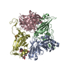 8becC 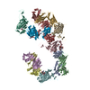 8bg1C 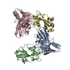 8bg3C 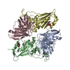 8bg4C 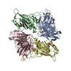 8bg5C 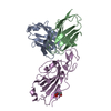 8bg6C 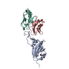 8bg8C 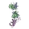 7k8mS  7kgjS S: Starting model for refinement C: citing same article ( |
|---|---|
| Similar structure data |


-Links
 PDBj
PDBj


- Assembly
| Deposited unit | 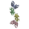
| ||||||||||||
|---|---|---|---|---|---|---|---|---|---|---|---|---|---|
| 1 | 
| ||||||||||||
| 2 | 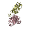
| ||||||||||||
| Unit cell |
|
-Components
| #1: Antibody | Mass: 26381.045 Da / Num. of mol.: 2 Source method: isolated from a genetically manipulated source Source: (gene. exp.) Homo sapiens (human) / Cell line (production host): S2 / Production host:  #2: Protein | Mass: 22361.924 Da / Num. of mol.: 2 Source method: isolated from a genetically manipulated source Source: (gene. exp.) Severe acute respiratory syndrome coronavirus 2Gene: S, 2 / Cell line (production host): S2 / Production host: #3: Sugar |   Type: D-saccharide, beta linking / Mass: 221.208 Da / Num. of mol.: 2 / Source method: obtained synthetically / Formula: C8H15NO6 Type: D-saccharide, beta linking / Mass: 221.208 Da / Num. of mol.: 2 / Source method: obtained synthetically / Formula: C8H15NO6#4: Water | ChemComp-HOH / |  Mass: 18.015 Da / Num. of mol.: 441 / Source method: isolated from a natural source / Formula: H2O Mass: 18.015 Da / Num. of mol.: 441 / Source method: isolated from a natural source / Formula: H2OHas ligand of interest | N | Has protein modification | Y | |
|---|
-Experimental details
-Experiment
| Experiment | Method: X-RAY DIFFRACTION / Number of used crystals: 1 |
|---|
- Sample preparation
| Crystal | Density Matthews: 3.01 Å3/Da / Density % sol: 59.17 % |
|---|---|
| Crystal grow | Temperature: 293 K / Method: vapor diffusion, sitting drop Details: pT1580-RBD at 9,8 mg/ml using 1.4 M Na-Tartrate, 100 mM Tris pH 8.5 as reservoir |
-Data collection
| Diffraction | Mean temperature: 100 K / Serial crystal experiment: N |
|---|---|
| Diffraction source | Source: SYNCHROTRON / Site: SLS  / Beamline: X06SA / Wavelength: 1 Å / Beamline: X06SA / Wavelength: 1 Å |
| Detector | Type: DECTRIS EIGER X 16M / Detector: PIXEL / Date: Mar 5, 2021 |
| Radiation | Protocol: SINGLE WAVELENGTH / Monochromatic (M) / Laue (L): M / Scattering type: x-ray |
| Radiation wavelength | Wavelength: 1 Å / Relative weight: 1 |
| Reflection | Resolution: 2.1→45.76 Å / Num. obs: 234717 / % possible obs: 94.11 % / Redundancy: 3.5 % / Biso Wilson estimate: 33.69 Å2 / CC1/2: 0.997 / CC star: 0.999 / Rmerge(I) obs: 0.09489 / Rpim(I) all: 0.05927 / Rrim(I) all: 0.1123 / Net I/σ(I): 9.19 |
| Reflection shell | Resolution: 2.1→2.175 Å / Redundancy: 3.6 % / Rmerge(I) obs: 1.21 / Mean I/σ(I) obs: 1.15 / Num. unique obs: 5914 / CC1/2: 0.444 / CC star: 0.784 / Rpim(I) all: 0.7422 / Rrim(I) all: 1.424 / % possible all: 88.15 |
- Processing
| Software |
| |||||||||||||||||||||||||||||||||||||||||||||||||||||||||||||||||||||||||||||||||||||||||||||||||||||||||||||||||||||||||||||
|---|---|---|---|---|---|---|---|---|---|---|---|---|---|---|---|---|---|---|---|---|---|---|---|---|---|---|---|---|---|---|---|---|---|---|---|---|---|---|---|---|---|---|---|---|---|---|---|---|---|---|---|---|---|---|---|---|---|---|---|---|---|---|---|---|---|---|---|---|---|---|---|---|---|---|---|---|---|---|---|---|---|---|---|---|---|---|---|---|---|---|---|---|---|---|---|---|---|---|---|---|---|---|---|---|---|---|---|---|---|---|---|---|---|---|---|---|---|---|---|---|---|---|---|---|---|---|
| Refinement | Method to determine structure: MOLECULAR REPLACEMENT Starting model: 7KGJ, 7K8M Resolution: 2.1→45.76 Å / SU ML: 0.2757 / Cross valid method: FREE R-VALUE / σ(F): 0.07 / Phase error: 25.3512 Stereochemistry target values: GeoStd + Monomer Library + CDL v1.2
| |||||||||||||||||||||||||||||||||||||||||||||||||||||||||||||||||||||||||||||||||||||||||||||||||||||||||||||||||||||||||||||
| Solvent computation | Shrinkage radii: 0.9 Å / VDW probe radii: 1.11 Å / Solvent model: FLAT BULK SOLVENT MODEL | |||||||||||||||||||||||||||||||||||||||||||||||||||||||||||||||||||||||||||||||||||||||||||||||||||||||||||||||||||||||||||||
| Displacement parameters | Biso mean: 40.44 Å2 | |||||||||||||||||||||||||||||||||||||||||||||||||||||||||||||||||||||||||||||||||||||||||||||||||||||||||||||||||||||||||||||
| Refinement step | Cycle: LAST / Resolution: 2.1→45.76 Å
| |||||||||||||||||||||||||||||||||||||||||||||||||||||||||||||||||||||||||||||||||||||||||||||||||||||||||||||||||||||||||||||
| Refine LS restraints |
| |||||||||||||||||||||||||||||||||||||||||||||||||||||||||||||||||||||||||||||||||||||||||||||||||||||||||||||||||||||||||||||
| LS refinement shell |
| |||||||||||||||||||||||||||||||||||||||||||||||||||||||||||||||||||||||||||||||||||||||||||||||||||||||||||||||||||||||||||||
| Refinement TLS params. | Method: refined / Refine-ID: X-RAY DIFFRACTION
| |||||||||||||||||||||||||||||||||||||||||||||||||||||||||||||||||||||||||||||||||||||||||||||||||||||||||||||||||||||||||||||
| Refinement TLS group | Refine-ID: X-RAY DIFFRACTION
|
