 Movie
Movie Controller

[English] 日本語
 Yorodumi
- PDB-8bbj: Secretagogin (mouse) in complex with its target peptide from Synt... -
+ Open data

- Basic information
| Entry | Database: PDB / ID: 8bbj | ||||||
|---|---|---|---|---|---|---|---|
| Title | Secretagogin (mouse) in complex with its target peptide from Syntaxin-4 | ||||||
 Components Components |
| ||||||
 Keywords Keywords | PEPTIDE BINDING PROTEIN / Calcium dependent / protein complex | ||||||
| Function / homology |  Function and homology information Function and homology informationDisinhibition of SNARE formation / sphingomyelin phosphodiesterase activator activity / trans-Golgi Network Vesicle Budding / Other interleukin signaling / cornified envelope assembly / positive regulation of eosinophil degranulation / neuron projection membrane / myelin sheath adaxonal region / storage vacuole / lateral loop ...Disinhibition of SNARE formation / sphingomyelin phosphodiesterase activator activity / trans-Golgi Network Vesicle Budding / Other interleukin signaling / cornified envelope assembly / positive regulation of eosinophil degranulation / neuron projection membrane / myelin sheath adaxonal region / storage vacuole / lateral loop / specific granule / SNARE complex / SNAP receptor activity / positive regulation of chemotaxis / stereocilium / positive regulation of protein localization to cell surface / regulation of extrinsic apoptotic signaling pathway via death domain receptors / ER-Phagosome pathway / protein localization to cell surface / positive regulation of immunoglobulin production / transport vesicle membrane / SNARE complex assembly / synaptic vesicle exocytosis / positive regulation of insulin secretion involved in cellular response to glucose stimulus / regulation of postsynaptic membrane neurotransmitter receptor levels / phagocytic vesicle / positive regulation of cell adhesion / bioluminescence / SNARE binding / positive regulation of protein localization to plasma membrane / generation of precursor metabolites and energy / intracellular protein transport / trans-Golgi network / sensory perception of sound / cellular response to type II interferon / Schaffer collateral - CA1 synapse / long-term synaptic potentiation / lamellipodium / presynapse / cellular response to oxidative stress / basolateral plasma membrane / dendritic spine / endosome / postsynapse / positive regulation of cell migration / positive regulation of cell population proliferation / calcium ion binding / perinuclear region of cytoplasm / glutamatergic synapse / cell surface / extracellular space / extracellular region / plasma membrane / cytoplasm Similarity search - Function | ||||||
| Biological species |   Aequorea victoria (jellyfish) Aequorea victoria (jellyfish) | ||||||
| Method |  X-RAY DIFFRACTION / SYNCHROTRON / MOLECULAR REPLACEMENT / Resolution: 2.65 Å X-RAY DIFFRACTION / SYNCHROTRON / MOLECULAR REPLACEMENT / Resolution: 2.65 Å | ||||||
 Authors Authors | Schnell, R. / Szodorai, E. | ||||||
| Funding support |  Denmark, 1items Denmark, 1items
| ||||||
 Citation Citation | Journal: Proc.Natl.Acad.Sci.USA / Year: 2024 Title: A hydrophobic groove in secretagogin allows for alternate interactions with SNAP-25 and syntaxin-4 in endocrine tissues. Authors: Szodorai, E. / Hevesi, Z. / Wagner, L. / Hokfelt, T.G.M. / Harkany, T. / Schnell, R. | ||||||
| History |
|
- Structure visualization
| Structure viewer | Molecule: MolmilJmol/JSmol |
|---|
- Downloads & links
-Download
| PDBx/mmCIF format | 8bbj.cif.gz | 186.5 KB | Display | PDBx/mmCIF format |
|---|---|---|---|---|
| PDB format | pdb8bbj.ent.gz | 145.8 KB | Display | PDB format |
| PDBx/mmJSON format | 8bbj.json.gz | Tree view | PDBx/mmJSON format | |
| Others |  Other downloads Other downloads |
-Validation report
| Arichive directory | https://data.pdbj.org/pub/pdb/validation_reports/bb/8bbjftp://data.pdbj.org/pub/pdb/validation_reports/bb/8bbj | HTTPS FTP |
|---|
-Related structure data
| Related structure data | 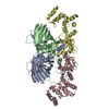 8banSC 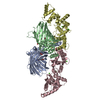 8bavC  1emaS S: Starting model for refinement C: citing same article ( |
|---|---|
| Similar structure data |
-Links
 PDBj
PDBj


- Assembly
| Deposited unit | 
| ||||||||
|---|---|---|---|---|---|---|---|---|---|
| 1 | 
| ||||||||
| 2 | 
| ||||||||
| Unit cell |
|
-Components
| #1: Protein | Mass: 29433.234 Da / Num. of mol.: 2 / Mutation: F64L,Q80R,I167T Source method: isolated from a genetically manipulated source Details: GFP-fusion protein carrying a short peptide from mouse Syntaxin-4 (sequence: GLQNLREEIKQLGREVRAQLKAIEPQKE) on its C-terminus. The Chromophore of GFP is formed from residues Ser65-Tyr66- ...Details: GFP-fusion protein carrying a short peptide from mouse Syntaxin-4 (sequence: GLQNLREEIKQLGREVRAQLKAIEPQKE) on its C-terminus. The Chromophore of GFP is formed from residues Ser65-Tyr66-Gly67, and represented as non-peptide residue CRO (Chromophore / Fluorophore). This alteration results in an apparent mismatch when comparing the encoded polypeptide sequence (present in databases) and the residues present in the PDB file. Source: (gene. exp.) Aequorea victoria (jellyfish), (gene. exp.) Gene: GFP, Stx4, Stx4a / Production host:  #2: Protein | Mass: 21998.104 Da / Num. of mol.: 2 Source method: isolated from a genetically manipulated source Source: (gene. exp.) #3: Chemical | ChemComp-CA /   Mass: 40.078 Da / Num. of mol.: 8 / Source method: obtained synthetically / Formula: Ca Mass: 40.078 Da / Num. of mol.: 8 / Source method: obtained synthetically / Formula: Ca#4: Chemical | ChemComp-CAC / |   Mass: 136.989 Da / Num. of mol.: 1 / Source method: obtained synthetically / Formula: C2H6AsO2 Mass: 136.989 Da / Num. of mol.: 1 / Source method: obtained synthetically / Formula: C2H6AsO2#5: Water | ChemComp-HOH / |  Mass: 18.015 Da / Num. of mol.: 12 / Source method: isolated from a natural source / Formula: H2O Mass: 18.015 Da / Num. of mol.: 12 / Source method: isolated from a natural source / Formula: H2OHas ligand of interest | N | Has protein modification | Y | |
|---|
-Experimental details
-Experiment
| Experiment | Method: X-RAY DIFFRACTION / Number of used crystals: 1 |
|---|
- Sample preparation
| Crystal | Density Matthews: 4.55 Å3/Da / Density % sol: 72 % / Description: rod |
|---|---|
| Crystal grow | Temperature: 277 K / Method: vapor diffusion / pH: 8 Details: 0.1 M Na cacodylate pH 5.5, 0.2 M NH4SO4, 25% v/v PEGSM (PEG-smear) |
-Data collection
| Diffraction | Mean temperature: 110 K / Serial crystal experiment: N |
|---|---|
| Diffraction source | Source: SYNCHROTRON / Site: MAX IV  / Beamline: BioMAX / Wavelength: 0.96863 Å / Beamline: BioMAX / Wavelength: 0.96863 Å |
| Detector | Type: DECTRIS PILATUS3 6M / Detector: PIXEL / Date: Feb 11, 2021 / Details: mirrors |
| Radiation | Monochromator: Si (111) / Protocol: SINGLE WAVELENGTH / Monochromatic (M) / Laue (L): M / Scattering type: x-ray |
| Radiation wavelength | Wavelength: 0.96863 Å / Relative weight: 1 |
| Reflection | Resolution: 2.65→78.68 Å / Num. obs: 28954 / % possible obs: 98.2 % / Redundancy: 3.3 % / Biso Wilson estimate: 60.4 Å2 / CC1/2: 0.998 / Rmerge(I) obs: 0.07 / Rpim(I) all: 0.059 / Net I/σ(I): 8.2 |
| Reflection shell | Resolution: 2.65→2.78 Å / Rmerge(I) obs: 0.678 / Mean I/σ(I) obs: 1.4 / Num. unique obs: 3847 / CC1/2: 0.598 / Rpim(I) all: 0.574 |
- Processing
| Software |
| ||||||||||||||||||||||||||||||||||||||||||||||||||||||||||||||||||||||||||||||||||||
|---|---|---|---|---|---|---|---|---|---|---|---|---|---|---|---|---|---|---|---|---|---|---|---|---|---|---|---|---|---|---|---|---|---|---|---|---|---|---|---|---|---|---|---|---|---|---|---|---|---|---|---|---|---|---|---|---|---|---|---|---|---|---|---|---|---|---|---|---|---|---|---|---|---|---|---|---|---|---|---|---|---|---|---|---|---|
| Refinement | Method to determine structure: MOLECULAR REPLACEMENT Starting model: 1EMA, 8BAN Resolution: 2.65→48.289 Å / SU ML: 0.35 / Cross valid method: THROUGHOUT / σ(F): 1.34 / Phase error: 31 / Stereochemistry target values: ML
| ||||||||||||||||||||||||||||||||||||||||||||||||||||||||||||||||||||||||||||||||||||
| Solvent computation | Shrinkage radii: 0.9 Å / VDW probe radii: 1.11 Å / Solvent model: FLAT BULK SOLVENT MODEL | ||||||||||||||||||||||||||||||||||||||||||||||||||||||||||||||||||||||||||||||||||||
| Refinement step | Cycle: LAST / Resolution: 2.65→48.289 Å
| ||||||||||||||||||||||||||||||||||||||||||||||||||||||||||||||||||||||||||||||||||||
| Refine LS restraints |
| ||||||||||||||||||||||||||||||||||||||||||||||||||||||||||||||||||||||||||||||||||||
| LS refinement shell |
|
