[English] 日本語
 Yorodumi
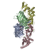
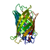
- PDB-8ban: Secretagogin (mouse) in complex with its target peptide from SNAP-25 -
+ Open data
- Basic information
| Entry | Database: PDB / ID: 8ban | ||||||
|---|---|---|---|---|---|---|---|
| Title | Secretagogin (mouse) in complex with its target peptide from SNAP-25 | ||||||
 Components Components |
| ||||||
 Keywords Keywords | PEPTIDE BINDING PROTEIN / Calcium-dependent / protein complex | ||||||
| Function / homology |  Function and homology information Function and homology informationToxicity of botulinum toxin type C (botC) / neurotransmitter uptake / Toxicity of botulinum toxin type E (botE) / GABA synthesis, release, reuptake and degradation / synaptic vesicle fusion to presynaptic active zone membrane / Acetylcholine Neurotransmitter Release Cycle / Toxicity of botulinum toxin type A (botA) / presynaptic dense core vesicle exocytosis / synaptobrevin 2-SNAP-25-syntaxin-1a-complexin I complex / Serotonin Neurotransmitter Release Cycle ...Toxicity of botulinum toxin type C (botC) / neurotransmitter uptake / Toxicity of botulinum toxin type E (botE) / GABA synthesis, release, reuptake and degradation / synaptic vesicle fusion to presynaptic active zone membrane / Acetylcholine Neurotransmitter Release Cycle / Toxicity of botulinum toxin type A (botA) / presynaptic dense core vesicle exocytosis / synaptobrevin 2-SNAP-25-syntaxin-1a-complexin I complex / Serotonin Neurotransmitter Release Cycle / synaptic vesicle docking / Norepinephrine Neurotransmitter Release Cycle / Dopamine Neurotransmitter Release Cycle / ribbon synapse / Glutamate Neurotransmitter Release Cycle / SNARE complex / SNAP receptor activity / Sensory processing of sound by inner hair cells of the cochlea / syntaxin-1 binding / Other interleukin signaling / transport vesicle membrane / regulation of neuron projection development / synaptic vesicle priming / exocytosis / tertiary granule membrane / associative learning / synaptic vesicle exocytosis / voltage-gated potassium channel activity / specific granule membrane / somatodendritic compartment / photoreceptor inner segment / regulation of insulin secretion / bioluminescence / generation of precursor metabolites and energy / Regulation of insulin secretion / locomotory behavior / trans-Golgi network / long-term synaptic potentiation / calcium-dependent protein binding / synaptic vesicle / growth cone / presynaptic membrane / cell cortex / chemical synaptic transmission / cytoskeleton / neuron projection / calcium ion binding / Neutrophil degranulation / lipid binding / perinuclear region of cytoplasm / glutamatergic synapse / extracellular region / membrane / plasma membrane / cytosol / cytoplasm Similarity search - Function | ||||||
| Biological species |   Homo sapiens (human) Homo sapiens (human) | ||||||
| Method |  X-RAY DIFFRACTION / X-RAY DIFFRACTION /  SYNCHROTRON / SYNCHROTRON /  MOLECULAR REPLACEMENT / Resolution: 2.35 Å MOLECULAR REPLACEMENT / Resolution: 2.35 Å | ||||||
 Authors Authors | Schnell, R. / Szodorai, E. | ||||||
| Funding support |  Denmark, 1items Denmark, 1items
| ||||||
 Citation Citation |  Journal: Proc.Natl.Acad.Sci.USA / Year: 2024 Journal: Proc.Natl.Acad.Sci.USA / Year: 2024Title: A hydrophobic groove in secretagogin allows for alternate interactions with SNAP-25 and syntaxin-4 in endocrine tissues. Authors: Szodorai, E. / Hevesi, Z. / Wagner, L. / Hokfelt, T.G.M. / Harkany, T. / Schnell, R. | ||||||
| History |
|
- Structure visualization
| Structure viewer | Molecule:  Molmil Molmil Jmol/JSmol Jmol/JSmol |
|---|
- Downloads & links
- Download
| PDBx/mmCIF format |  8ban.cif.gz 8ban.cif.gz | 221.2 KB | Display |  PDBx/mmCIF format PDBx/mmCIF format |
|---|---|---|---|---|
| PDB format |  pdb8ban.ent.gz pdb8ban.ent.gz | 174.5 KB | Display |  PDB format PDB format |
| PDBx/mmJSON format |  8ban.json.gz 8ban.json.gz | Tree view |  PDBx/mmJSON format PDBx/mmJSON format | |
| Others |  Other downloads Other downloads |
-Validation report
| Arichive directory |  https://data.pdbj.org/pub/pdb/validation_reports/ba/8ban https://data.pdbj.org/pub/pdb/validation_reports/ba/8ban ftp://data.pdbj.org/pub/pdb/validation_reports/ba/8ban ftp://data.pdbj.org/pub/pdb/validation_reports/ba/8ban | HTTPS FTP |
|---|
-Related structure data
| Related structure data |  8bavC  8bbjC  1emaS S: Starting model for refinement C: citing same article ( |
|---|---|
| Similar structure data | Similarity search - Function & homology  F&H Search F&H Search |
- Links
Links
- Assembly
| Deposited unit | 
| ||||||||
|---|---|---|---|---|---|---|---|---|---|
| 1 | 
| ||||||||
| 2 | 
| ||||||||
| Unit cell |
|
- Components
| #1: Protein | Mass: 28980.629 Da / Num. of mol.: 2 Mutation: F64L,Q80R,I167T MUTATIONS IN ENHANCED GFP (HIGHER FLUORESCENCE INTENSITY) Source method: isolated from a genetically manipulated source Details: GFP-fusion protein carrying a short peptide from human SNAP25 (sequence: GIIGNLRHMALDMGNEIDTQNRQID) on its C-terminus. The Chromophore of GFP is formed from residues Ser65-Tyr66-Gly67, and ...Details: GFP-fusion protein carrying a short peptide from human SNAP25 (sequence: GIIGNLRHMALDMGNEIDTQNRQID) on its C-terminus. The Chromophore of GFP is formed from residues Ser65-Tyr66-Gly67, and represented as non-peptide residue CRO (Chromophore / Fluorophore). This alteration results in an apparent mismatch when comparing the encoded polypeptide sequence (present in databases) and the residues present in the PDB file. Source: (gene. exp.)   Homo sapiens (human) Homo sapiens (human)Gene: GFP, SNAP25, SNAP / Production host:  #2: Protein | Mass: 32190.730 Da / Num. of mol.: 2 Source method: isolated from a genetically manipulated source Source: (gene. exp.)   #3: Chemical | ChemComp-CA / #4: Water | ChemComp-HOH / | Has ligand of interest | N | Has protein modification | Y | |
|---|
-Experimental details
-Experiment
| Experiment | Method:  X-RAY DIFFRACTION / Number of used crystals: 1 X-RAY DIFFRACTION / Number of used crystals: 1 |
|---|
- Sample preparation
| Crystal | Density Matthews: 2.24 Å3/Da / Density % sol: 45.11 % / Description: rod |
|---|---|
| Crystal grow | Temperature: 277 K / Method: vapor diffusion / pH: 6.5 Details: PEG 3350 (20%), 2 mM CaCl2, 0.1 M Bis-Tris-Propane pH 6.5, 25 mM Tris-HCl pH 8.0, 0.2 M NaI |
-Data collection
| Diffraction | Mean temperature: 110 K / Serial crystal experiment: N |
|---|---|
| Diffraction source | Source:  SYNCHROTRON / Site: SYNCHROTRON / Site:  MAX IV MAX IV  / Beamline: BioMAX / Wavelength: 0.97625 Å / Beamline: BioMAX / Wavelength: 0.97625 Å |
| Detector | Type: DECTRIS EIGER X 16M / Detector: PIXEL / Date: Feb 6, 2020 / Details: Toroidal mirrors |
| Radiation | Monochromator: Si(111) / Protocol: SINGLE WAVELENGTH / Monochromatic (M) / Laue (L): M / Scattering type: x-ray |
| Radiation wavelength | Wavelength: 0.97625 Å / Relative weight: 1 |
| Reflection | Resolution: 2.35→87.42 Å / Num. obs: 46202 / % possible obs: 99.2 % / Observed criterion σ(I): 2.1 / Redundancy: 7.3 % / CC1/2: 0.998 / Rmerge(I) obs: 0.098 / Net I/σ(I): 11.9 |
| Reflection shell | Resolution: 2.35→2.43 Å / Redundancy: 5.9 % / Rmerge(I) obs: 0.841 / Mean I/σ(I) obs: 2.1 / Num. unique obs: 4186 / CC1/2: 0.72 / Rpim(I) all: 0.529 / % possible all: 93 |
- Processing
| Software |
| ||||||||||||||||||||||||||||||||||||||||||||||||||||||||||||||||||||||||||||||||||||||||||||||||||||||||||||||||||||||||||||||||||||||||||||||||||||||||||||||||||||||||||||||||||||||
|---|---|---|---|---|---|---|---|---|---|---|---|---|---|---|---|---|---|---|---|---|---|---|---|---|---|---|---|---|---|---|---|---|---|---|---|---|---|---|---|---|---|---|---|---|---|---|---|---|---|---|---|---|---|---|---|---|---|---|---|---|---|---|---|---|---|---|---|---|---|---|---|---|---|---|---|---|---|---|---|---|---|---|---|---|---|---|---|---|---|---|---|---|---|---|---|---|---|---|---|---|---|---|---|---|---|---|---|---|---|---|---|---|---|---|---|---|---|---|---|---|---|---|---|---|---|---|---|---|---|---|---|---|---|---|---|---|---|---|---|---|---|---|---|---|---|---|---|---|---|---|---|---|---|---|---|---|---|---|---|---|---|---|---|---|---|---|---|---|---|---|---|---|---|---|---|---|---|---|---|---|---|---|---|
| Refinement | Method to determine structure:  MOLECULAR REPLACEMENT MOLECULAR REPLACEMENTStarting model: 1EMA Resolution: 2.35→87.42 Å / Cor.coef. Fo:Fc: 0.954 / Cor.coef. Fo:Fc free: 0.921 / SU B: 8.787 / SU ML: 0.202 / Cross valid method: THROUGHOUT / ESU R: 0.422 / ESU R Free: 0.261 / Stereochemistry target values: MAXIMUM LIKELIHOOD / Details: HYDROGENS HAVE BEEN ADDED IN THE RIDING POSITIONS
| ||||||||||||||||||||||||||||||||||||||||||||||||||||||||||||||||||||||||||||||||||||||||||||||||||||||||||||||||||||||||||||||||||||||||||||||||||||||||||||||||||||||||||||||||||||||
| Solvent computation | Ion probe radii: 0.8 Å / Shrinkage radii: 0.8 Å / VDW probe radii: 1.2 Å / Solvent model: MASK | ||||||||||||||||||||||||||||||||||||||||||||||||||||||||||||||||||||||||||||||||||||||||||||||||||||||||||||||||||||||||||||||||||||||||||||||||||||||||||||||||||||||||||||||||||||||
| Displacement parameters | Biso mean: 48.655 Å2
| ||||||||||||||||||||||||||||||||||||||||||||||||||||||||||||||||||||||||||||||||||||||||||||||||||||||||||||||||||||||||||||||||||||||||||||||||||||||||||||||||||||||||||||||||||||||
| Refinement step | Cycle: 1 / Resolution: 2.35→87.42 Å
| ||||||||||||||||||||||||||||||||||||||||||||||||||||||||||||||||||||||||||||||||||||||||||||||||||||||||||||||||||||||||||||||||||||||||||||||||||||||||||||||||||||||||||||||||||||||
| Refine LS restraints |
|
 Movie
Movie Controller


 PDBj
PDBj


