 Movie
Movie Controller

[English] 日本語
 Yorodumi
- PDB-8b6j: Cryo-EM structure of cytochrome bc1 complex (complex-III) from re... -
+ Open data

- Basic information
| Entry | Database: PDB / ID: 8b6j | |||||||||||||||
|---|---|---|---|---|---|---|---|---|---|---|---|---|---|---|---|---|
| Title | Cryo-EM structure of cytochrome bc1 complex (complex-III) from respiratory supercomplex of Tetrahymena thermophila | |||||||||||||||
 Components Components |
| |||||||||||||||
 Keywords Keywords | ELECTRON TRANSPORT / Ciliate / mitochondrial / cytochrome bc1 / supercomplex | |||||||||||||||
| Function / homology |  Function and homology information Function and homology informationquinol-cytochrome-c reductase / mitochondrial electron transport, ubiquinol to cytochrome c / respiratory electron transport chain / 2 iron, 2 sulfur cluster binding / metallopeptidase activity / electron transfer activity / oxidoreductase activity / mitochondrial inner membrane / heme binding / mitochondrion ...quinol-cytochrome-c reductase / mitochondrial electron transport, ubiquinol to cytochrome c / respiratory electron transport chain / 2 iron, 2 sulfur cluster binding / metallopeptidase activity / electron transfer activity / oxidoreductase activity / mitochondrial inner membrane / heme binding / mitochondrion / metal ion binding / membrane Similarity search - Function | |||||||||||||||
| Biological species |  Tetrahymena thermophila SB210 (eukaryote) Tetrahymena thermophila SB210 (eukaryote) | |||||||||||||||
| Method | ELECTRON MICROSCOPY / single particle reconstruction / cryo EM / Resolution: 2.8 Å | |||||||||||||||
 Authors Authors | Muhleip, A. / Kock Flygaard, R. / Amunts, A. | |||||||||||||||
| Funding support |  Sweden, European Union, 4items Sweden, European Union, 4items
| |||||||||||||||
 Citation Citation | Journal: Nature / Year: 2023 Title: Structural basis of mitochondrial membrane bending by the I-II-III-IV supercomplex. Authors: Alexander Mühleip / Rasmus Kock Flygaard / Rozbeh Baradaran / Outi Haapanen / Thomas Gruhl / Victor Tobiasson / Amandine Maréchal / Vivek Sharma / Alexey Amunts /     Abstract: Mitochondrial energy conversion requires an intricate architecture of the inner mitochondrial membrane. Here we show that a supercomplex containing all four respiratory chain components contributes ...Mitochondrial energy conversion requires an intricate architecture of the inner mitochondrial membrane. Here we show that a supercomplex containing all four respiratory chain components contributes to membrane curvature induction in ciliates. We report cryo-electron microscopy and cryo-tomography structures of the supercomplex that comprises 150 different proteins and 311 bound lipids, forming a stable 5.8-MDa assembly. Owing to subunit acquisition and extension, complex I associates with a complex IV dimer, generating a wedge-shaped gap that serves as a binding site for complex II. Together with a tilted complex III dimer association, it results in a curved membrane region. Using molecular dynamics simulations, we demonstrate that the divergent supercomplex actively contributes to the membrane curvature induction and tubulation of cristae. Our findings highlight how the evolution of protein subunits of respiratory complexes has led to the I-II-III-IV supercomplex that contributes to the shaping of the bioenergetic membrane, thereby enabling its functional specialization. #1: Journal: Biorxiv / Year: 2022Title: Structural basis of mitochondrial membrane bending by I-II-III2-IV2 supercomplex Authors: Muhleip, A. / Flygaard, R.K. / Haapanen, O. / Baradaran, R. / Gruhl, T. / Tobiasson, V. / Marechal, A. / Sharma, V. / Amunts, A. | |||||||||||||||
| History |
|
- Structure visualization
| Structure viewer | Molecule: MolmilJmol/JSmol |
|---|
- Downloads & links
-Download
| PDBx/mmCIF format | 8b6j.cif.gz | 1.9 MB | Display | PDBx/mmCIF format |
|---|---|---|---|---|
| PDB format | pdb8b6j.ent.gz | 1.6 MB | Display | PDB format |
| PDBx/mmJSON format | 8b6j.json.gz | Tree view | PDBx/mmJSON format | |
| Others |  Other downloads Other downloads |
-Validation report
| Summary document | 8b6j_validation.pdf.gz | 3.1 MB | Display | wwPDB validaton report |
|---|---|---|---|---|
| Full document | 8b6j_full_validation.pdf.gz | 3.2 MB | Display | |
| Data in XML | 8b6j_validation.xml.gz | 159.1 KB | Display | |
| Data in CIF | 8b6j_validation.cif.gz | 230.2 KB | Display | |
| Arichive directory | https://data.pdbj.org/pub/pdb/validation_reports/b6/8b6jftp://data.pdbj.org/pub/pdb/validation_reports/b6/8b6j | HTTPS FTP |
-Related structure data
| Related structure data |  15868MC  8b6fC  8b6gC 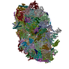 8b6hC  8bqsC M: map data used to model this data C: citing same article ( |
|---|---|
| Similar structure data |


-Links
 PDBj
PDBj


- Assembly
| Deposited unit | 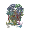
|
|---|---|
| 1 |
|
-Components
-Protein , 8 types, 16 molecules AaBbCcDdEeFfGgJj
| #1: Protein | Mass: 58023.914 Da / Num. of mol.: 2 / Source method: isolated from a natural source / Source: (natural) Tetrahymena thermophila SB210 (eukaryote) / Strain: SB210 / References: UniProt: I7MGU2#2: Protein | Mass: 53030.543 Da / Num. of mol.: 2 / Source method: isolated from a natural source / Source: (natural) Tetrahymena thermophila SB210 (eukaryote) / Strain: SB210 / References: UniProt: I7MJ25#3: Protein | Mass: 50635.547 Da / Num. of mol.: 2 / Source method: isolated from a natural source / Source: (natural) Tetrahymena thermophila SB210 (eukaryote) / References: UniProt: Q950Z1#4: Protein | Mass: 37616.629 Da / Num. of mol.: 2 / Source method: isolated from a natural source / Source: (natural) Tetrahymena thermophila SB210 (eukaryote) / Strain: SB210 / References: UniProt: Q24IM5#5: Protein | Mass: 30696.961 Da / Num. of mol.: 2 / Source method: isolated from a natural source / Source: (natural) Tetrahymena thermophila SB210 (eukaryote) / Strain: SB210 / References: UniProt: I7MIC7#6: Protein | Mass: 9885.511 Da / Num. of mol.: 2 / Source method: isolated from a natural source / Source: (natural) Tetrahymena thermophila SB210 (eukaryote) / Strain: SB210 / References: UniProt: Q23K66#7: Protein | Mass: 39193.523 Da / Num. of mol.: 2 / Source method: isolated from a natural source / Source: (natural) Tetrahymena thermophila SB210 (eukaryote) / Strain: SB210 / References: UniProt: Q23F81#10: Protein | Mass: 5634.938 Da / Num. of mol.: 2 / Source method: isolated from a natural source / Source: (natural) Tetrahymena thermophila SB210 (eukaryote) |
|---|
-Transmembrane protein, ... , 3 types, 6 molecules HhIiKk
| #8: Protein | Mass: 15677.771 Da / Num. of mol.: 2 / Source method: isolated from a natural source / Source: (natural) Tetrahymena thermophila SB210 (eukaryote) / Strain: SB210 / References: UniProt: I7M484#9: Protein | Mass: 14262.554 Da / Num. of mol.: 2 / Source method: isolated from a natural source / Source: (natural) Tetrahymena thermophila SB210 (eukaryote) / Strain: SB210 / References: UniProt: I7MM45#11: Protein | Mass: 7455.741 Da / Num. of mol.: 2 / Source method: isolated from a natural source / Source: (natural) Tetrahymena thermophila SB210 (eukaryote) / Strain: SB210 / References: UniProt: I7MFL6 |
|---|
-Protein/peptide , 1 types, 2 molecules lL
| #12: Protein/peptide | Mass: 4850.612 Da / Num. of mol.: 2 / Source method: isolated from a natural source / Source: (natural) Tetrahymena thermophila SB210 (eukaryote) |
|---|
-Non-polymers , 7 types, 43 molecules 


| #13: Chemical | ChemComp-PC1 /  Mass: 790.145 Da / Num. of mol.: 15 / Source method: obtained synthetically / Formula: C44H88NO8P / Comment: phospholipid*YM Mass: 790.145 Da / Num. of mol.: 15 / Source method: obtained synthetically / Formula: C44H88NO8P / Comment: phospholipid*YM#14: Chemical | ChemComp-HEM /  Mass: 616.487 Da / Num. of mol.: 4 / Source method: obtained synthetically / Formula: C34H32FeN4O4 / Feature type: SUBJECT OF INVESTIGATION Mass: 616.487 Da / Num. of mol.: 4 / Source method: obtained synthetically / Formula: C34H32FeN4O4 / Feature type: SUBJECT OF INVESTIGATION#15: Chemical | ChemComp-CDL /  Mass: 1464.043 Da / Num. of mol.: 15 / Source method: obtained synthetically / Formula: C81H156O17P2 / Feature type: SUBJECT OF INVESTIGATION / Comment: phospholipid*YM Mass: 1464.043 Da / Num. of mol.: 15 / Source method: obtained synthetically / Formula: C81H156O17P2 / Feature type: SUBJECT OF INVESTIGATION / Comment: phospholipid*YM#16: Chemical |  Mass: 727.109 Da / Num. of mol.: 3 / Source method: obtained synthetically / Formula: C49H74O4 / Feature type: SUBJECT OF INVESTIGATION Mass: 727.109 Da / Num. of mol.: 3 / Source method: obtained synthetically / Formula: C49H74O4 / Feature type: SUBJECT OF INVESTIGATION#17: Chemical |  Mass: 744.034 Da / Num. of mol.: 2 / Source method: obtained synthetically / Formula: C41H78NO8P / Comment: DOPE, phospholipid*YM Mass: 744.034 Da / Num. of mol.: 2 / Source method: obtained synthetically / Formula: C41H78NO8P / Comment: DOPE, phospholipid*YM#18: Chemical |  Mass: 618.503 Da / Num. of mol.: 2 / Source method: obtained synthetically / Formula: C34H34FeN4O4 / Feature type: SUBJECT OF INVESTIGATION Mass: 618.503 Da / Num. of mol.: 2 / Source method: obtained synthetically / Formula: C34H34FeN4O4 / Feature type: SUBJECT OF INVESTIGATION#19: Chemical |  Mass: 175.820 Da / Num. of mol.: 2 / Source method: obtained synthetically / Formula: Fe2S2 / Feature type: SUBJECT OF INVESTIGATION Mass: 175.820 Da / Num. of mol.: 2 / Source method: obtained synthetically / Formula: Fe2S2 / Feature type: SUBJECT OF INVESTIGATION |
|---|
-Details
| Has ligand of interest | Y |
|---|
-Experimental details
-Experiment
| Experiment | Method: ELECTRON MICROSCOPY |
|---|---|
| EM experiment | Aggregation state: PARTICLE / 3D reconstruction method: single particle reconstruction |
- Sample preparation
| Component | Name: Dimeric cytochrome bc1 complex (complex-III2) / Type: COMPLEX / Entity ID: #1-#12 / Source: NATURAL |
|---|---|
| Molecular weight | Value: 0.64 MDa / Experimental value: NO |
| Source (natural) | Organism:  Tetrahymena thermophila (eukaryote) / Organelle: Mitochondrion Tetrahymena thermophila (eukaryote) / Organelle: Mitochondrion |
| Buffer solution | pH: 7.5 |
| Specimen | Embedding applied: NO / Shadowing applied: NO / Staining applied: NO / Vitrification applied: YES |
| Vitrification | Cryogen name: ETHANE |
- Electron microscopy imaging
| Experimental equipment |  Model: Titan Krios / Image courtesy: FEI Company |
|---|---|
| Microscopy | Model: FEI TITAN KRIOS |
| Electron gun | Electron source:  FIELD EMISSION GUN / Accelerating voltage: 300 kV / Illumination mode: FLOOD BEAM FIELD EMISSION GUN / Accelerating voltage: 300 kV / Illumination mode: FLOOD BEAM |
| Electron lens | Mode: BRIGHT FIELD / Nominal defocus max: 2600 nm / Nominal defocus min: 600 nm |
| Image recording | Electron dose: 25.66 e/Å2 / Film or detector model: GATAN K2 QUANTUM (4k x 4k) |
- Processing
| CTF correction | Type: PHASE FLIPPING AND AMPLITUDE CORRECTION |
|---|---|
| 3D reconstruction | Resolution: 2.8 Å / Resolution method: FSC 0.143 CUT-OFF / Num. of particles: 138746 / Symmetry type: POINT |
