 Movie
Movie Controller

[English] 日本語
 Yorodumi
- PDB-8aol: Crystal structure of S-layer protein SlpX from Lactobacillus acid... -
+ Open data

- Basic information
| Entry | Database: PDB / ID: 8aol | ||||||
|---|---|---|---|---|---|---|---|
| Title | Crystal structure of S-layer protein SlpX from Lactobacillus acidophilus, domain III (aa 363-499) | ||||||
 Components Components | SlpX | ||||||
 Keywords Keywords | CELL ADHESION / Surface Layer / SlpX / Cell wall binding | ||||||
| Function / homology | Surface layer protein A domain / Surface layer protein A domain / metal ion binding / ACETATE ION / DI(HYDROXYETHYL)ETHER / PHOSPHATE ION / SlpX Function and homology information Function and homology information | ||||||
| Biological species |  Lactobacillus acidophilus ATCC 4796 (bacteria) Lactobacillus acidophilus ATCC 4796 (bacteria) | ||||||
| Method |  X-RAY DIFFRACTION / SYNCHROTRON / MOLECULAR REPLACEMENT / Resolution: 2.4 Å X-RAY DIFFRACTION / SYNCHROTRON / MOLECULAR REPLACEMENT / Resolution: 2.4 Å | ||||||
 Authors Authors | Sagmeister, T. / Damisch, E. / Eder, M. / Dordic, A. / Vejzovic, D. / Pavkov-Keller, T. | ||||||
| Funding support |  Austria, 1items Austria, 1items
| ||||||
 Citation Citation | Journal: Proc.Natl.Acad.Sci.USA / Year: 2024 Title: The molecular architecture of Lactobacillus S-layer: Assembly and attachment to teichoic acids. Authors: Sagmeister, T. / Gubensak, N. / Buhlheller, C. / Grininger, C. / Eder, M. / Ðordic, A. / Millan, C. / Medina, A. / Murcia, P.A.S. / Berni, F. / Hynonen, U. / Vejzovic, D. / Damisch, E. ...Authors: Sagmeister, T. / Gubensak, N. / Buhlheller, C. / Grininger, C. / Eder, M. / Ðordic, A. / Millan, C. / Medina, A. / Murcia, P.A.S. / Berni, F. / Hynonen, U. / Vejzovic, D. / Damisch, E. / Kulminskaya, N. / Petrowitsch, L. / Oberer, M. / Palva, A. / Malanovic, N. / Codee, J. / Keller, W. / Uson, I. / Pavkov-Keller, T. | ||||||
| History |
|
- Structure visualization
| Structure viewer | Molecule: MolmilJmol/JSmol |
|---|
- Downloads & links
-Download
| PDBx/mmCIF format | 8aol.cif.gz | 76.8 KB | Display | PDBx/mmCIF format |
|---|---|---|---|---|
| PDB format | pdb8aol.ent.gz | 54.3 KB | Display | PDB format |
| PDBx/mmJSON format | 8aol.json.gz | Tree view | PDBx/mmJSON format | |
| Others |  Other downloads Other downloads |
-Validation report
| Arichive directory | https://data.pdbj.org/pub/pdb/validation_reports/ao/8aolftp://data.pdbj.org/pub/pdb/validation_reports/ao/8aol | HTTPS FTP |
|---|
-Related structure data
| Related structure data |  7qecC  7qehC 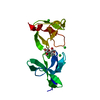 7qfgC  7qfiC  7qfjC  7qfkC 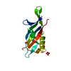 7qflC  7qldC  7qleC  7qlhC  8aluSC  8bt9C  8q1oC S: Starting model for refinement C: citing same article ( |
|---|---|
| Similar structure data |
-Links
 PDBj
PDBj- Assembly
| Deposited unit | 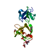
| |||||||||
|---|---|---|---|---|---|---|---|---|---|---|
| 1 |
| |||||||||
| Unit cell |
| |||||||||
| Components on special symmetry positions |
|
-Components
-Protein , 1 types, 1 molecules A
| #1: Protein | Mass: 17032.344 Da / Num. of mol.: 1 Source method: isolated from a genetically manipulated source Source: (gene. exp.) Lactobacillus acidophilus ATCC 4796 (bacteria)Gene: slpX, LBA0512 / Production host: |
|---|
-Non-polymers , 6 types, 152 molecules 


| #2: Chemical |  Mass: 65.409 Da / Num. of mol.: 3 / Source method: obtained synthetically / Formula: Zn Mass: 65.409 Da / Num. of mol.: 3 / Source method: obtained synthetically / Formula: Zn#3: Chemical |  Mass: 94.971 Da / Num. of mol.: 2 / Source method: obtained synthetically / Formula: PO4 Mass: 94.971 Da / Num. of mol.: 2 / Source method: obtained synthetically / Formula: PO4#4: Chemical | ChemComp-CA / |  Mass: 40.078 Da / Num. of mol.: 1 / Source method: obtained synthetically / Formula: Ca Mass: 40.078 Da / Num. of mol.: 1 / Source method: obtained synthetically / Formula: Ca#5: Chemical |  Mass: 59.044 Da / Num. of mol.: 2 / Source method: obtained synthetically / Formula: C2H3O2 Mass: 59.044 Da / Num. of mol.: 2 / Source method: obtained synthetically / Formula: C2H3O2#6: Chemical | ChemComp-PEG / |  Mass: 106.120 Da / Num. of mol.: 1 / Source method: obtained synthetically / Formula: C4H10O3 Mass: 106.120 Da / Num. of mol.: 1 / Source method: obtained synthetically / Formula: C4H10O3#7: Water | ChemComp-HOH / | Mass: 18.015 Da / Num. of mol.: 143 / Source method: isolated from a natural source / Formula: H2O |
|---|
-Details
| Has ligand of interest | N |
|---|---|
| Has protein modification | N |
-Experimental details
-Experiment
| Experiment | Method: X-RAY DIFFRACTION / Number of used crystals: 1 |
|---|
- Sample preparation
| Crystal | Density Matthews: 5.3 Å3/Da / Density % sol: 76.81 % |
|---|---|
| Crystal grow | Temperature: 293.15 K / Method: vapor diffusion, sitting drop Details: Protein stock solution of 10 mg/mL in 20 mM Hepes pH 8 and 100 mM NaCl; Wizard 1/2 screen condition 66 (20 % w/v PEG 3000, 200 mM Ca(OAc)2, 100 mM Tris base/HCl pH 7.0) with protein end ...Details: Protein stock solution of 10 mg/mL in 20 mM Hepes pH 8 and 100 mM NaCl; Wizard 1/2 screen condition 66 (20 % w/v PEG 3000, 200 mM Ca(OAc)2, 100 mM Tris base/HCl pH 7.0) with protein end concentration of 5 mg/mL corresponding to 50% of protein solution in the 1.0 uL drop |
-Data collection
| Diffraction | Mean temperature: 100 K / Serial crystal experiment: N | |||||||||||||||||||||
|---|---|---|---|---|---|---|---|---|---|---|---|---|---|---|---|---|---|---|---|---|---|---|
| Diffraction source | Source: SYNCHROTRON / Site: ESRF  / Beamline: ID23-1 / Wavelength: 1.0332 Å / Beamline: ID23-1 / Wavelength: 1.0332 Å | |||||||||||||||||||||
| Detector | Type: DECTRIS PILATUS 6M / Detector: PIXEL / Date: Jul 22, 2018 | |||||||||||||||||||||
| Radiation | Protocol: SINGLE WAVELENGTH / Monochromatic (M) / Laue (L): M / Scattering type: x-ray | |||||||||||||||||||||
| Radiation wavelength | Wavelength: 1.0332 Å / Relative weight: 1 | |||||||||||||||||||||
| Reflection | Resolution: 2.4→48.432 Å / Num. obs: 14935 / % possible obs: 100 % / Redundancy: 19.1 % / CC1/2: 0.997 / Rmerge(I) obs: 0.198 / Rpim(I) all: 0.065 / Rrim(I) all: 0.209 / Net I/σ(I): 10.1 | |||||||||||||||||||||
| Reflection shell | Diffraction-ID: 1
|
- Processing
| Software |
| |||||||||||||||||||||||||||||||||||||||||||||||||||||||||||||||||||||||||||||||||||||||||||||||||||||||||||||||||||||||||||||||||||||||||||||||||||||||||||||||||||||||||||||||||||||||||||||||||||||||||||||||||||||||||||||||||||||||
|---|---|---|---|---|---|---|---|---|---|---|---|---|---|---|---|---|---|---|---|---|---|---|---|---|---|---|---|---|---|---|---|---|---|---|---|---|---|---|---|---|---|---|---|---|---|---|---|---|---|---|---|---|---|---|---|---|---|---|---|---|---|---|---|---|---|---|---|---|---|---|---|---|---|---|---|---|---|---|---|---|---|---|---|---|---|---|---|---|---|---|---|---|---|---|---|---|---|---|---|---|---|---|---|---|---|---|---|---|---|---|---|---|---|---|---|---|---|---|---|---|---|---|---|---|---|---|---|---|---|---|---|---|---|---|---|---|---|---|---|---|---|---|---|---|---|---|---|---|---|---|---|---|---|---|---|---|---|---|---|---|---|---|---|---|---|---|---|---|---|---|---|---|---|---|---|---|---|---|---|---|---|---|---|---|---|---|---|---|---|---|---|---|---|---|---|---|---|---|---|---|---|---|---|---|---|---|---|---|---|---|---|---|---|---|---|---|---|---|---|---|---|---|---|---|---|---|---|---|---|---|---|---|
| Refinement | Method to determine structure: MOLECULAR REPLACEMENT Starting model: 8ALU Resolution: 2.4→48.432 Å / Cor.coef. Fo:Fc: 0.968 / Cor.coef. Fo:Fc free: 0.951 / WRfactor Rfree: 0.197 / WRfactor Rwork: 0.17 / SU B: 6.849 / SU ML: 0.138 / Average fsc free: 0.8733 / Average fsc work: 0.8785 / Cross valid method: FREE R-VALUE / ESU R: 0.168 / ESU R Free: 0.153 Details: Hydrogens have been added in their riding positions
| |||||||||||||||||||||||||||||||||||||||||||||||||||||||||||||||||||||||||||||||||||||||||||||||||||||||||||||||||||||||||||||||||||||||||||||||||||||||||||||||||||||||||||||||||||||||||||||||||||||||||||||||||||||||||||||||||||||||
| Solvent computation | Ion probe radii: 0.8 Å / Shrinkage radii: 0.8 Å / VDW probe radii: 1.2 Å / Solvent model: MASK BULK SOLVENT | |||||||||||||||||||||||||||||||||||||||||||||||||||||||||||||||||||||||||||||||||||||||||||||||||||||||||||||||||||||||||||||||||||||||||||||||||||||||||||||||||||||||||||||||||||||||||||||||||||||||||||||||||||||||||||||||||||||||
| Displacement parameters | Biso mean: 46.79 Å2
| |||||||||||||||||||||||||||||||||||||||||||||||||||||||||||||||||||||||||||||||||||||||||||||||||||||||||||||||||||||||||||||||||||||||||||||||||||||||||||||||||||||||||||||||||||||||||||||||||||||||||||||||||||||||||||||||||||||||
| Refinement step | Cycle: LAST / Resolution: 2.4→48.432 Å
| |||||||||||||||||||||||||||||||||||||||||||||||||||||||||||||||||||||||||||||||||||||||||||||||||||||||||||||||||||||||||||||||||||||||||||||||||||||||||||||||||||||||||||||||||||||||||||||||||||||||||||||||||||||||||||||||||||||||
| Refine LS restraints |
| |||||||||||||||||||||||||||||||||||||||||||||||||||||||||||||||||||||||||||||||||||||||||||||||||||||||||||||||||||||||||||||||||||||||||||||||||||||||||||||||||||||||||||||||||||||||||||||||||||||||||||||||||||||||||||||||||||||||
| LS refinement shell | Refine-ID: X-RAY DIFFRACTION / Total num. of bins used: 20
|
