 Movie
Movie Controller

[English] 日本語
 Yorodumi
- PDB-8aed: Broadly neutralizing DARPin bnD.9 in complex with the HIV-1 envel... -
+ Open data

- Basic information
| Entry | Database: PDB / ID: 8aed | ||||||
|---|---|---|---|---|---|---|---|
| Title | Broadly neutralizing DARPin bnD.9 in complex with the HIV-1 envelope variable loop 3 peptide V3 (BG505) | ||||||
 Components Components |
| ||||||
 Keywords Keywords | DE NOVO PROTEIN / DARPin / HIV | ||||||
| Function / homology |  Function and homology information Function and homology informationsymbiont-mediated perturbation of host defense response / positive regulation of plasma membrane raft polarization / positive regulation of receptor clustering / host cell endosome membrane / clathrin-dependent endocytosis of virus by host cell / viral protein processing / fusion of virus membrane with host plasma membrane / fusion of virus membrane with host endosome membrane / viral envelope / virion attachment to host cell ...symbiont-mediated perturbation of host defense response / positive regulation of plasma membrane raft polarization / positive regulation of receptor clustering / host cell endosome membrane / clathrin-dependent endocytosis of virus by host cell / viral protein processing / fusion of virus membrane with host plasma membrane / fusion of virus membrane with host endosome membrane / viral envelope / virion attachment to host cell / host cell plasma membrane / virion membrane / structural molecule activity / membrane Similarity search - Function | ||||||
| Biological species | synthetic construct (others)  Human immunodeficiency virus 1 Human immunodeficiency virus 1 | ||||||
| Method |  X-RAY DIFFRACTION / SYNCHROTRON / MOLECULAR REPLACEMENT / Resolution: 1.17 Å X-RAY DIFFRACTION / SYNCHROTRON / MOLECULAR REPLACEMENT / Resolution: 1.17 Å | ||||||
 Authors Authors | Mittl, P. / Gloegl, M. | ||||||
| Funding support |  Switzerland, 1items Switzerland, 1items
| ||||||
 Citation Citation | Journal: Nat Struct Mol Biol / Year: 2023 Title: Trapping the HIV-1 V3 loop in a helical conformation enables broad neutralization. Authors: Matthias Glögl / Nikolas Friedrich / Gabriele Cerutti / Thomas Lemmin / Young D Kwon / Jason Gorman / Liridona Maliqi / Peer R E Mittl / Maria C Hesselman / Daniel Schmidt / Jacqueline ...Authors: Matthias Glögl / Nikolas Friedrich / Gabriele Cerutti / Thomas Lemmin / Young D Kwon / Jason Gorman / Liridona Maliqi / Peer R E Mittl / Maria C Hesselman / Daniel Schmidt / Jacqueline Weber / Caio Foulkes / Adam S Dingens / Tatsiana Bylund / Adam S Olia / Raffaello Verardi / Thomas Reinberg / Nicolas S Baumann / Peter Rusert / Birgit Dreier / Lawrence Shapiro / Peter D Kwong / Andreas Plückthun / Alexandra Trkola /  Abstract: The third variable (V3) loop on the human immunodeficiency virus 1 (HIV-1) envelope glycoprotein trimer is indispensable for virus cell entry. Conformational masking of V3 within the trimer allows ...The third variable (V3) loop on the human immunodeficiency virus 1 (HIV-1) envelope glycoprotein trimer is indispensable for virus cell entry. Conformational masking of V3 within the trimer allows efficient neutralization via V3 only by rare, broadly neutralizing glycan-dependent antibodies targeting the closed prefusion trimer but not by abundant antibodies that access the V3 crown on open trimers after CD4 attachment. Here, we report on a distinct category of V3-specific inhibitors based on designed ankyrin repeat protein (DARPin) technology that reinstitute the CD4-bound state as a key neutralization target with up to >90% breadth. Broadly neutralizing DARPins (bnDs) bound V3 solely on open envelope and recognized a four-turn amphipathic α-helix in the carboxy-terminal half of V3 (amino acids 314-324), which we termed 'αV3C'. The bnD contact surface on αV3C was as conserved as the CD4 binding site. Molecular dynamics and escape mutation analyses underscored the functional relevance of αV3C, highlighting the potential of αV3C-based inhibitors and, more generally, of postattachment inhibition of HIV-1. | ||||||
| History |
|
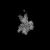
- Structure visualization
| Structure viewer | Molecule: MolmilJmol/JSmol |
|---|
- Downloads & links
-Download
| PDBx/mmCIF format | 8aed.cif.gz | 225.1 KB | Display | PDBx/mmCIF format |
|---|---|---|---|---|
| PDB format | pdb8aed.ent.gz | 184.2 KB | Display | PDB format |
| PDBx/mmJSON format | 8aed.json.gz | Tree view | PDBx/mmJSON format | |
| Others |  Other downloads Other downloads |
-Validation report
| Arichive directory | https://data.pdbj.org/pub/pdb/validation_reports/ae/8aedftp://data.pdbj.org/pub/pdb/validation_reports/ae/8aed | HTTPS FTP |
|---|
-Related structure data
| Related structure data |  7txdC  7z7cC  2qyjS C: citing same article ( S: Starting model for refinement |
|---|---|
| Similar structure data |
-Links
 PDBj
PDBj


- Assembly
| Deposited unit | 
| ||||||||
|---|---|---|---|---|---|---|---|---|---|
| 1 | 
| ||||||||
| 2 | 
| ||||||||
| Unit cell |
|
-Components
-Protein / Protein/peptide , 2 types, 4 molecules ABCD
| #1: Protein | Mass: 17211.521 Da / Num. of mol.: 2 Source method: isolated from a genetically manipulated source Source: (gene. exp.) synthetic construct (others) / Production host:  #2: Protein/peptide | Mass: 2566.889 Da / Num. of mol.: 2 / Source method: obtained synthetically / Source: (synth.) Human immunodeficiency virus 1 / References: UniProt: Q9Q714 |
|---|
-Non-polymers , 5 types, 504 molecules 


| #3: Chemical | ChemComp-EDO /  Mass: 62.068 Da / Num. of mol.: 4 / Source method: obtained synthetically / Formula: C2H6O2 Mass: 62.068 Da / Num. of mol.: 4 / Source method: obtained synthetically / Formula: C2H6O2#4: Chemical |  Mass: 59.044 Da / Num. of mol.: 2 / Source method: obtained synthetically / Formula: C2H3O2 Mass: 59.044 Da / Num. of mol.: 2 / Source method: obtained synthetically / Formula: C2H3O2#5: Chemical |  Mass: 24.305 Da / Num. of mol.: 3 / Source method: obtained synthetically / Formula: Mg Mass: 24.305 Da / Num. of mol.: 3 / Source method: obtained synthetically / Formula: Mg#6: Chemical |  Mass: 192.124 Da / Num. of mol.: 2 / Source method: obtained synthetically / Formula: C6H8O7 Mass: 192.124 Da / Num. of mol.: 2 / Source method: obtained synthetically / Formula: C6H8O7#7: Water | ChemComp-HOH / | Mass: 18.015 Da / Num. of mol.: 493 / Source method: isolated from a natural source / Formula: H2O |
|---|
-Details
| Has ligand of interest | N |
|---|---|
| Has protein modification | N |
-Experimental details
-Experiment
| Experiment | Method: X-RAY DIFFRACTION / Number of used crystals: 1 |
|---|
- Sample preparation
| Crystal | Density Matthews: 2.04 Å3/Da / Density % sol: 39.56 % |
|---|---|
| Crystal grow | Temperature: 293 K / Method: vapor diffusion, sitting drop Details: 100 mM trisodium citrate, pH 4.31 165 mM magnesium acetate 20% PEG smear broad (PSB) |
-Data collection
| Diffraction | Mean temperature: 100 K / Serial crystal experiment: N |
|---|---|
| Diffraction source | Source: SYNCHROTRON / Site: SLS / Beamline: X06DA / Wavelength: 1 Å |
| Detector | Type: DECTRIS PILATUS 12M / Detector: PIXEL / Date: Nov 25, 2019 |
| Radiation | Protocol: SINGLE WAVELENGTH / Monochromatic (M) / Laue (L): M / Scattering type: x-ray |
| Radiation wavelength | Wavelength: 1 Å / Relative weight: 1 |
| Reflection | Resolution: 1.17→57.39 Å / Num. obs: 79970 / % possible obs: 87.5 % / Redundancy: 6.2 % / Biso Wilson estimate: 10.56 Å2 / CC1/2: 0.994 / Rmerge(I) obs: 0.071 / Rpim(I) all: 0.046 / Rrim(I) all: 0.085 / Net I/σ(I): 12.3 |
| Reflection shell | Resolution: 1.17→1.25 Å / Mean I/σ(I) obs: 1.8 / Num. unique obs: 3995 / CC1/2: 0.635 / % possible all: 43.1 |
- Processing
| Software |
| ||||||||||||||||||||||||||||||||||||||||||||||||||||||||||||||||||||||||||||||||||||||||||||||||||||||||||||||||||||||||||||||||||||||||||||||||||||||||||||||||||||||||||||||||||||||||||||||||||||||||||||||||||
|---|---|---|---|---|---|---|---|---|---|---|---|---|---|---|---|---|---|---|---|---|---|---|---|---|---|---|---|---|---|---|---|---|---|---|---|---|---|---|---|---|---|---|---|---|---|---|---|---|---|---|---|---|---|---|---|---|---|---|---|---|---|---|---|---|---|---|---|---|---|---|---|---|---|---|---|---|---|---|---|---|---|---|---|---|---|---|---|---|---|---|---|---|---|---|---|---|---|---|---|---|---|---|---|---|---|---|---|---|---|---|---|---|---|---|---|---|---|---|---|---|---|---|---|---|---|---|---|---|---|---|---|---|---|---|---|---|---|---|---|---|---|---|---|---|---|---|---|---|---|---|---|---|---|---|---|---|---|---|---|---|---|---|---|---|---|---|---|---|---|---|---|---|---|---|---|---|---|---|---|---|---|---|---|---|---|---|---|---|---|---|---|---|---|---|---|---|---|---|---|---|---|---|---|---|---|---|---|---|---|---|---|
| Refinement | Method to determine structure: MOLECULAR REPLACEMENT Starting model: 2QYJ Resolution: 1.17→50.23 Å / SU ML: 0.1 / Cross valid method: THROUGHOUT / σ(F): 1.34 / Phase error: 20.12 / Stereochemistry target values: ML
| ||||||||||||||||||||||||||||||||||||||||||||||||||||||||||||||||||||||||||||||||||||||||||||||||||||||||||||||||||||||||||||||||||||||||||||||||||||||||||||||||||||||||||||||||||||||||||||||||||||||||||||||||||
| Solvent computation | Shrinkage radii: 0.9 Å / VDW probe radii: 1.11 Å / Solvent model: FLAT BULK SOLVENT MODEL | ||||||||||||||||||||||||||||||||||||||||||||||||||||||||||||||||||||||||||||||||||||||||||||||||||||||||||||||||||||||||||||||||||||||||||||||||||||||||||||||||||||||||||||||||||||||||||||||||||||||||||||||||||
| Displacement parameters | Biso max: 69.1 Å2 / Biso mean: 15.9323 Å2 / Biso min: 5.25 Å2 | ||||||||||||||||||||||||||||||||||||||||||||||||||||||||||||||||||||||||||||||||||||||||||||||||||||||||||||||||||||||||||||||||||||||||||||||||||||||||||||||||||||||||||||||||||||||||||||||||||||||||||||||||||
| Refinement step | Cycle: final / Resolution: 1.17→50.23 Å
| ||||||||||||||||||||||||||||||||||||||||||||||||||||||||||||||||||||||||||||||||||||||||||||||||||||||||||||||||||||||||||||||||||||||||||||||||||||||||||||||||||||||||||||||||||||||||||||||||||||||||||||||||||
| LS refinement shell | Refine-ID: X-RAY DIFFRACTION / Rfactor Rfree error: 0 / Total num. of bins used: 29
|
