 Movie
Movie Controller

[English] 日本語
 Yorodumi
- PDB-8a8t: Malonyl-CoA reductase from Chloroflexus aurantiacus - C-terminal ... -
+ Open data

- Basic information
| Entry | Database: PDB / ID: 8a8t | ||||||||||||
|---|---|---|---|---|---|---|---|---|---|---|---|---|---|
| Title | Malonyl-CoA reductase from Chloroflexus aurantiacus - C-terminal NADP and malonate bound | ||||||||||||
 Components Components | Short-chain dehydrogenase/reductase SDR | ||||||||||||
 Keywords Keywords | OXIDOREDUCTASE / Malonyl-CoA / Chloroflexus aurantiacus / reductase / 3-hydroxypropionate / 3-HP cycle / bi-functional enzyme / carbon fixation | ||||||||||||
| Function / homology |  Function and homology information Function and homology informationfatty acid elongation / oxidoreductase activity, acting on the CH-OH group of donors, NAD or NADP as acceptor / nucleotide binding / metal ion binding Similarity search - Function | ||||||||||||
| Biological species |   Chloroflexus aurantiacus J-10-fl (bacteria) Chloroflexus aurantiacus J-10-fl (bacteria) | ||||||||||||
| Method |  X-RAY DIFFRACTION / SYNCHROTRON / MOLECULAR REPLACEMENT / Resolution: 2.11 Å X-RAY DIFFRACTION / SYNCHROTRON / MOLECULAR REPLACEMENT / Resolution: 2.11 Å | ||||||||||||
 Authors Authors | Kabasakal, B.V. / Murray, J.W. | ||||||||||||
| Funding support |  United Kingdom, 3items United Kingdom, 3items
| ||||||||||||
 Citation Citation | Journal: Biochimie / Year: 2023 Title: Dynamic lid domain of Chloroflexus aurantiacus Malonyl-CoA reductase controls the reaction. Authors: Kabasakal, B.V. / Cotton, C.A.R. / Murray, J.W. #1: Journal: Biorxiv / Year: 2023Title: Dynamic lid domain of Chloroflexus aurantiacus Malonyl-CoA Reductase controls the reaction Authors: Kabasakal, B.V. / Cotton, C.A.R. / Murray, J.W. | ||||||||||||
| History |
|
- Structure visualization
| Structure viewer | Molecule: MolmilJmol/JSmol |
|---|
- Downloads & links
-Download
| PDBx/mmCIF format | 8a8t.cif.gz | 272.9 KB | Display | PDBx/mmCIF format |
|---|---|---|---|---|
| PDB format | pdb8a8t.ent.gz | 215.8 KB | Display | PDB format |
| PDBx/mmJSON format | 8a8t.json.gz | Tree view | PDBx/mmJSON format | |
| Others |  Other downloads Other downloads |
-Validation report
| Summary document | 8a8t_validation.pdf.gz | 1.1 MB | Display | wwPDB validaton report |
|---|---|---|---|---|
| Full document | 8a8t_full_validation.pdf.gz | 1.1 MB | Display | |
| Data in XML | 8a8t_validation.xml.gz | 26.4 KB | Display | |
| Data in CIF | 8a8t_validation.cif.gz | 38.4 KB | Display | |
| Arichive directory | https://data.pdbj.org/pub/pdb/validation_reports/a8/8a8tftp://data.pdbj.org/pub/pdb/validation_reports/a8/8a8t | HTTPS FTP |
-Related structure data
| Related structure data |  8a30SC  8a7sC  8aeoC  8aeqC 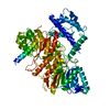 8aerC 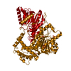 8aetC 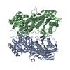 8aewC S: Starting model for refinement C: citing same article ( |
|---|---|
| Similar structure data |
-Links
 PDBj
PDBj


- Assembly
| Deposited unit | 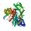
| ||||||||||||
|---|---|---|---|---|---|---|---|---|---|---|---|---|---|
| 1 | 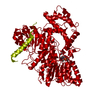
| ||||||||||||
| Unit cell |
| ||||||||||||
| Components on special symmetry positions |
|
-Components
| #1: Protein | Mass: 73321.453 Da / Num. of mol.: 1 Source method: isolated from a genetically manipulated source Source: (gene. exp.) Chloroflexus aurantiacus J-10-fl (bacteria)Gene: Caur_2614 / Production host: |
|---|---|
| #2: Chemical | ChemComp-NAP /   Mass: 743.405 Da / Num. of mol.: 1 / Source method: obtained synthetically / Formula: C21H28N7O17P3 / Feature type: SUBJECT OF INVESTIGATION Mass: 743.405 Da / Num. of mol.: 1 / Source method: obtained synthetically / Formula: C21H28N7O17P3 / Feature type: SUBJECT OF INVESTIGATION |
| #3: Chemical | ChemComp-MLI /   Mass: 102.046 Da / Num. of mol.: 1 / Source method: obtained synthetically / Formula: C3H2O4 / Feature type: SUBJECT OF INVESTIGATION Mass: 102.046 Da / Num. of mol.: 1 / Source method: obtained synthetically / Formula: C3H2O4 / Feature type: SUBJECT OF INVESTIGATION |
| #4: Water | ChemComp-HOH /  Mass: 18.015 Da / Num. of mol.: 282 / Source method: isolated from a natural source / Formula: H2O Mass: 18.015 Da / Num. of mol.: 282 / Source method: isolated from a natural source / Formula: H2O |
| Has ligand of interest | Y |
-Experimental details
-Experiment
| Experiment | Method: X-RAY DIFFRACTION / Number of used crystals: 1 |
|---|
- Sample preparation
| Crystal | Density Matthews: 3.12 Å3/Da / Density % sol: 60.62 % |
|---|---|
| Crystal grow | Temperature: 293 K / Method: vapor diffusion, hanging drop / pH: 7 Details: 0.8 M succinic acid, pH 7.0, 0.5 mM NADPH co-crystallised |
-Data collection
| Diffraction | Mean temperature: 100 K / Serial crystal experiment: N |
|---|---|
| Diffraction source | Source: SYNCHROTRON / Site: Diamond / Beamline: I04 / Wavelength: 0.97949 Å |
| Detector | Type: DECTRIS PILATUS 6M-F / Detector: PIXEL / Date: Feb 29, 2016 |
| Radiation | Protocol: SINGLE WAVELENGTH / Monochromatic (M) / Laue (L): M / Scattering type: x-ray |
| Radiation wavelength | Wavelength: 0.97949 Å / Relative weight: 1 |
| Reflection | Resolution: 2.11→63.15 Å / Num. obs: 100168 / % possible obs: 98.2 % / Redundancy: 1.9 % / Biso Wilson estimate: 42.78 Å2 / CC1/2: 0.998 / CC star: 1 / Rmerge(I) obs: 0.03785 / Rpim(I) all: 0.03785 / Rrim(I) all: 0.05353 / Net I/σ(I): 11.14 |
| Reflection shell | Resolution: 2.11→2.185 Å / Redundancy: 2 % / Rmerge(I) obs: 0.5348 / Mean I/σ(I) obs: 1.44 / Num. unique obs: 5221 / CC1/2: 0.624 / CC star: 0.877 / Rpim(I) all: 0.5348 / Rrim(I) all: 0.7564 / % possible all: 99.87 |
- Processing
| Software |
| |||||||||||||||||||||||||||||||||||||||||||||||||||||||||||||||||||||||||||||||||||||||||||||||||||||||||||||||||||||||||||||||||||||||||||||||||||||||||||||||||||||||||||||||||||||||||||||||||||||||||||||||||||||||||
|---|---|---|---|---|---|---|---|---|---|---|---|---|---|---|---|---|---|---|---|---|---|---|---|---|---|---|---|---|---|---|---|---|---|---|---|---|---|---|---|---|---|---|---|---|---|---|---|---|---|---|---|---|---|---|---|---|---|---|---|---|---|---|---|---|---|---|---|---|---|---|---|---|---|---|---|---|---|---|---|---|---|---|---|---|---|---|---|---|---|---|---|---|---|---|---|---|---|---|---|---|---|---|---|---|---|---|---|---|---|---|---|---|---|---|---|---|---|---|---|---|---|---|---|---|---|---|---|---|---|---|---|---|---|---|---|---|---|---|---|---|---|---|---|---|---|---|---|---|---|---|---|---|---|---|---|---|---|---|---|---|---|---|---|---|---|---|---|---|---|---|---|---|---|---|---|---|---|---|---|---|---|---|---|---|---|---|---|---|---|---|---|---|---|---|---|---|---|---|---|---|---|---|---|---|---|---|---|---|---|---|---|---|---|---|---|---|---|---|
| Refinement | Method to determine structure: MOLECULAR REPLACEMENT Starting model: 8A30 Resolution: 2.11→63.15 Å / SU ML: 0.3 / Cross valid method: FREE R-VALUE / σ(F): 0.02 / Phase error: 25.62 / Stereochemistry target values: ML
| |||||||||||||||||||||||||||||||||||||||||||||||||||||||||||||||||||||||||||||||||||||||||||||||||||||||||||||||||||||||||||||||||||||||||||||||||||||||||||||||||||||||||||||||||||||||||||||||||||||||||||||||||||||||||
| Solvent computation | Shrinkage radii: 0.9 Å / VDW probe radii: 1.11 Å / Solvent model: FLAT BULK SOLVENT MODEL | |||||||||||||||||||||||||||||||||||||||||||||||||||||||||||||||||||||||||||||||||||||||||||||||||||||||||||||||||||||||||||||||||||||||||||||||||||||||||||||||||||||||||||||||||||||||||||||||||||||||||||||||||||||||||
| Refinement step | Cycle: LAST / Resolution: 2.11→63.15 Å
| |||||||||||||||||||||||||||||||||||||||||||||||||||||||||||||||||||||||||||||||||||||||||||||||||||||||||||||||||||||||||||||||||||||||||||||||||||||||||||||||||||||||||||||||||||||||||||||||||||||||||||||||||||||||||
| Refine LS restraints |
| |||||||||||||||||||||||||||||||||||||||||||||||||||||||||||||||||||||||||||||||||||||||||||||||||||||||||||||||||||||||||||||||||||||||||||||||||||||||||||||||||||||||||||||||||||||||||||||||||||||||||||||||||||||||||
| LS refinement shell |
| |||||||||||||||||||||||||||||||||||||||||||||||||||||||||||||||||||||||||||||||||||||||||||||||||||||||||||||||||||||||||||||||||||||||||||||||||||||||||||||||||||||||||||||||||||||||||||||||||||||||||||||||||||||||||
| Refinement TLS params. | Method: refined / Refine-ID: X-RAY DIFFRACTION
| |||||||||||||||||||||||||||||||||||||||||||||||||||||||||||||||||||||||||||||||||||||||||||||||||||||||||||||||||||||||||||||||||||||||||||||||||||||||||||||||||||||||||||||||||||||||||||||||||||||||||||||||||||||||||
| Refinement TLS group |
|
