 Movie
Movie Controller

[English] 日本語
 Yorodumi
- PDB-7zvc: Second crystal form of the mature glutamic-class prolyl-endopepti... -
+ Open data

- Basic information
| Entry | Database: PDB / ID: 7zvc | ||||||
|---|---|---|---|---|---|---|---|
| Title | Second crystal form of the mature glutamic-class prolyl-endopeptidase neprosin at 1.85 A resolution. | ||||||
 Components Components |
| ||||||
 Keywords Keywords | HYDROLASE / glutamic endopeptidase / mature form / coeliac disease therapy / plant protease / pitcher plant | ||||||
| Function / homology | GLYCINE / NICKEL (II) ION Function and homology information Function and homology information | ||||||
| Biological species |  Nepenthes ventricosa x Nepenthes alata (plant) Nepenthes ventricosa x Nepenthes alata (plant) | ||||||
| Method |  X-RAY DIFFRACTION / SYNCHROTRON / MOLECULAR REPLACEMENT / Resolution: 1.85 Å X-RAY DIFFRACTION / SYNCHROTRON / MOLECULAR REPLACEMENT / Resolution: 1.85 Å | ||||||
 Authors Authors | Rodriguez-Banqueri, A. / Eckhard, U. / Del Amo-Maestro, L. / Mendes, S.R. / Guevara, T. / Gomis-Ruth, F.X. | ||||||
| Funding support | 1items
| ||||||
 Citation Citation | Journal: Nat Commun / Year: 2022 Title: Molecular and in vivo studies of a glutamate-class prolyl-endopeptidase for coeliac disease therapy. Authors: Del Amo-Maestro, L. / Mendes, S.R. / Rodriguez-Banqueri, A. / Garzon-Flores, L. / Girbal, M. / Rodriguez-Lagunas, M.J. / Guevara, T. / Franch, A. / Perez-Cano, F.J. / Eckhard, U. / Gomis-Ruth, F.X. | ||||||
| History |
|
- Structure visualization
| Structure viewer | Molecule: MolmilJmol/JSmol |
|---|
- Downloads & links
-Download
| PDBx/mmCIF format | 7zvc.cif.gz | 122.6 KB | Display | PDBx/mmCIF format |
|---|---|---|---|---|
| PDB format | pdb7zvc.ent.gz | 92.5 KB | Display | PDB format |
| PDBx/mmJSON format | 7zvc.json.gz | Tree view | PDBx/mmJSON format | |
| Others |  Other downloads Other downloads |
-Validation report
| Arichive directory | https://data.pdbj.org/pub/pdb/validation_reports/zv/7zvcftp://data.pdbj.org/pub/pdb/validation_reports/zv/7zvc | HTTPS FTP |
|---|
-Related structure data
| Related structure data |  7zu8SC 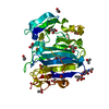 7zvaC  7zvbC S: Starting model for refinement C: citing same article ( |
|---|---|
| Similar structure data |
-Links
 PDBj
PDBj

- Assembly
| Deposited unit | 
| ||||||||
|---|---|---|---|---|---|---|---|---|---|
| 1 |
| ||||||||
| Unit cell |
|
-Components
-Protein / Protein/peptide , 2 types, 2 molecules AC
| #1: Protein | Mass: 28644.033 Da / Num. of mol.: 1 Source method: isolated from a genetically manipulated source Details: Mature form (S129-Q380) of the glutamic endopeptidase neprosin after autolytic removal of the N-terminal pro-domain at low pH. Of note, N-terminus after maturation was confirmed by Edman degradation. Source: (gene. exp.) Nepenthes ventricosa x Nepenthes alata (plant)Plasmid: pCMV-Sport 6 Cell (production host): Suspension-adapted 293 human embryonic kidney (HEK) cells Production host:  Homo sapiens (human) Homo sapiens (human) |
|---|---|
| #2: Protein/peptide | Mass: 246.222 Da / Num. of mol.: 1 Source method: isolated from a genetically manipulated source Source: (gene. exp.) Nepenthes ventricosa x Nepenthes alata (plant)Production host: Homo sapiens (human) |
-Sugars , 2 types, 2 molecules 
| #3: Polysaccharide | beta-D-mannopyranose-(1-4)-2-acetamido-2-deoxy-beta-D-glucopyranose-(1-4)-2-acetamido-2-deoxy-beta- ...beta-D-mannopyranose-(1-4)-2-acetamido-2-deoxy-beta-D-glucopyranose-(1-4)-2-acetamido-2-deoxy-beta-D-glucopyranose |
|---|---|
| #4: Sugar | ChemComp-NAG /  Type: D-saccharide, beta linking / Mass: 221.208 Da / Num. of mol.: 1 / Source method: obtained synthetically / Formula: C8H15NO6 Type: D-saccharide, beta linking / Mass: 221.208 Da / Num. of mol.: 1 / Source method: obtained synthetically / Formula: C8H15NO6 |
-Non-polymers , 4 types, 255 molecules 


| #5: Chemical |  Mass: 96.063 Da / Num. of mol.: 3 / Source method: isolated from a natural source / Formula: SO4 Mass: 96.063 Da / Num. of mol.: 3 / Source method: isolated from a natural source / Formula: SO4#6: Chemical | ChemComp-NI / |  Mass: 58.693 Da / Num. of mol.: 1 / Source method: obtained synthetically / Formula: Ni Mass: 58.693 Da / Num. of mol.: 1 / Source method: obtained synthetically / Formula: Ni#7: Chemical | ChemComp-GLY / |  Type: peptide linking / Mass: 75.067 Da / Num. of mol.: 1 / Source method: isolated from a natural source / Formula: C2H5NO2 Type: peptide linking / Mass: 75.067 Da / Num. of mol.: 1 / Source method: isolated from a natural source / Formula: C2H5NO2#8: Water | ChemComp-HOH / | Mass: 18.015 Da / Num. of mol.: 250 / Source method: isolated from a natural source / Formula: H2O |
|---|
-Details
| Has ligand of interest | N |
|---|---|
| Has protein modification | Y |
-Experimental details
-Experiment
| Experiment | Method: X-RAY DIFFRACTION / Number of used crystals: 1 |
|---|
- Sample preparation
| Crystal | Density Matthews: 2.56 Å3/Da / Density % sol: 51.89 % |
|---|---|
| Crystal grow | Temperature: 293 K / Method: vapor diffusion, sitting drop Details: 0.1 M sodium citrate tribasic pH 5.6, 0.5 M ammonium sulphate, 1 M lithium sulphate |
-Data collection
| Diffraction | Mean temperature: 100 K / Serial crystal experiment: N |
|---|---|
| Diffraction source | Source: SYNCHROTRON / Site: ALBA  / Beamline: XALOC / Wavelength: 0.97926 Å / Beamline: XALOC / Wavelength: 0.97926 Å |
| Detector | Type: DECTRIS PILATUS 6M / Detector: PIXEL / Date: Apr 24, 2022 |
| Radiation | Protocol: SINGLE WAVELENGTH / Monochromatic (M) / Laue (L): M / Scattering type: x-ray |
| Radiation wavelength | Wavelength: 0.97926 Å / Relative weight: 1 |
| Reflection | Resolution: 1.85→61.5 Å / Num. obs: 24437 / % possible obs: 97.1 % / Redundancy: 6.55 % / CC1/2: 0.998 / Net I/σ(I): 12.4 |
| Reflection shell | Resolution: 1.85→1.916 Å / Num. unique obs: 2007 / CC1/2: 0.729 |
- Processing
| Software |
| |||||||||||||||||||||||||||||||||||||||||||||||||||||||||||||||||||||||||||
|---|---|---|---|---|---|---|---|---|---|---|---|---|---|---|---|---|---|---|---|---|---|---|---|---|---|---|---|---|---|---|---|---|---|---|---|---|---|---|---|---|---|---|---|---|---|---|---|---|---|---|---|---|---|---|---|---|---|---|---|---|---|---|---|---|---|---|---|---|---|---|---|---|---|---|---|---|
| Refinement | Method to determine structure: MOLECULAR REPLACEMENT Starting model: 7ZU8 Resolution: 1.85→20.66 Å / Cor.coef. Fo:Fc: 0.947 / Cor.coef. Fo:Fc free: 0.939 / SU R Cruickshank DPI: 0.127 / Cross valid method: THROUGHOUT / SU R Blow DPI: 0.136 / SU Rfree Blow DPI: 0.121 / SU Rfree Cruickshank DPI: 0.117
| |||||||||||||||||||||||||||||||||||||||||||||||||||||||||||||||||||||||||||
| Displacement parameters | Biso mean: 27.86 Å2
| |||||||||||||||||||||||||||||||||||||||||||||||||||||||||||||||||||||||||||
| Refine analyze | Luzzati coordinate error obs: 0.22 Å | |||||||||||||||||||||||||||||||||||||||||||||||||||||||||||||||||||||||||||
| Refinement step | Cycle: LAST / Resolution: 1.85→20.66 Å
| |||||||||||||||||||||||||||||||||||||||||||||||||||||||||||||||||||||||||||
| Refine LS restraints |
| |||||||||||||||||||||||||||||||||||||||||||||||||||||||||||||||||||||||||||
| LS refinement shell | Resolution: 1.85→1.88 Å
| |||||||||||||||||||||||||||||||||||||||||||||||||||||||||||||||||||||||||||
| Refinement TLS params. | Refine-ID: X-RAY DIFFRACTION
| |||||||||||||||||||||||||||||||||||||||||||||||||||||||||||||||||||||||||||
| Refinement TLS group |
|
