 Movie
Movie Controller

[English] 日本語
 Yorodumi
- PDB-7zva: Crystal Structure of the native zymogen form of the glutamic-clas... -
+ Open data

- Basic information
| Entry | Database: PDB / ID: 7zva | ||||||
|---|---|---|---|---|---|---|---|
| Title | Crystal Structure of the native zymogen form of the glutamic-class prolyl-endopeptidase neprosin at 1.80 A resolution. | ||||||
 Components Components | C-terminal peptidase | ||||||
 Keywords Keywords | HYDROLASE / glutamic endopeptidase / zymogen / proform / coeliac disease therapy / plant protease | ||||||
| Function / homology | ACETATE ION / ISOPROPYL ALCOHOL Function and homology information Function and homology information | ||||||
| Biological species |  Nepenthes ventricosa x Nepenthes alata (plant) Nepenthes ventricosa x Nepenthes alata (plant) | ||||||
| Method |  X-RAY DIFFRACTION / SYNCHROTRON / MOLECULAR REPLACEMENT / Resolution: 1.8 Å X-RAY DIFFRACTION / SYNCHROTRON / MOLECULAR REPLACEMENT / Resolution: 1.8 Å | ||||||
 Authors Authors | Del Amo-Maestro, L. / Eckhard, U. / Rodriguez-Banqueri, A. / Mendes, S.R. / Guevara, T. / Gomis-Ruth, F.X. | ||||||
| Funding support | 1items
| ||||||
 Citation Citation | Journal: Nat Commun / Year: 2022 Title: Molecular and in vivo studies of a glutamate-class prolyl-endopeptidase for coeliac disease therapy. Authors: Del Amo-Maestro, L. / Mendes, S.R. / Rodriguez-Banqueri, A. / Garzon-Flores, L. / Girbal, M. / Rodriguez-Lagunas, M.J. / Guevara, T. / Franch, A. / Perez-Cano, F.J. / Eckhard, U. / Gomis-Ruth, F.X. | ||||||
| History |
|
- Structure visualization
| Structure viewer | Molecule: MolmilJmol/JSmol |
|---|
- Downloads & links
-Download
| PDBx/mmCIF format | 7zva.cif.gz | 154.4 KB | Display | PDBx/mmCIF format |
|---|---|---|---|---|
| PDB format | pdb7zva.ent.gz | 119.4 KB | Display | PDB format |
| PDBx/mmJSON format | 7zva.json.gz | Tree view | PDBx/mmJSON format | |
| Others |  Other downloads Other downloads |
-Validation report
| Arichive directory | https://data.pdbj.org/pub/pdb/validation_reports/zv/7zvaftp://data.pdbj.org/pub/pdb/validation_reports/zv/7zva | HTTPS FTP |
|---|
-Related structure data
| Related structure data |  7zu8SC  7zvbC  7zvcC S: Starting model for refinement C: citing same article ( |
|---|---|
| Similar structure data |
-Links
 PDBj
PDBj
- Assembly
| Deposited unit | 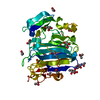
| ||||||||
|---|---|---|---|---|---|---|---|---|---|
| 1 |
| ||||||||
| Unit cell |
|
-Components
-Protein , 1 types, 1 molecules A
| #1: Protein | Mass: 43006.703 Da / Num. of mol.: 1 Source method: isolated from a genetically manipulated source Details: Pro-form of the glutamic endopeptidase neprosin (R25 to Q380) with an N-terminal IgK leader sequence and a C-terminal non-cleavable His6-tag. Source: (gene. exp.) Nepenthes ventricosa x Nepenthes alata (plant)Plasmid: pCMV-Sport 6 Cell line (production host): Suspension-adapted 293 human embryonic kidney (HEK) cells Production host:  Homo sapiens (human) Homo sapiens (human) |
|---|
-Sugars , 2 types, 2 molecules
| #2: Polysaccharide | alpha-L-fucopyranose-(1-6)-2-acetamido-2-deoxy-beta-D-glucopyranose Source method: isolated from a genetically manipulated source |
|---|---|
| #3: Polysaccharide | 2-acetamido-2-deoxy-beta-D-glucopyranose-(1-4)-2-acetamido-2-deoxy-beta-D-glucopyranose Source method: isolated from a genetically manipulated source |
-Non-polymers , 4 types, 270 molecules 


| #4: Chemical | ChemComp-GOL /  Mass: 92.094 Da / Num. of mol.: 5 / Source method: obtained synthetically / Formula: C3H8O3 Mass: 92.094 Da / Num. of mol.: 5 / Source method: obtained synthetically / Formula: C3H8O3#5: Chemical | ChemComp-ACT /  Mass: 59.044 Da / Num. of mol.: 8 / Source method: obtained synthetically / Formula: C2H3O2 Mass: 59.044 Da / Num. of mol.: 8 / Source method: obtained synthetically / Formula: C2H3O2#6: Chemical | ChemComp-IPA / |  Mass: 60.095 Da / Num. of mol.: 1 / Source method: obtained synthetically / Formula: C3H8O Mass: 60.095 Da / Num. of mol.: 1 / Source method: obtained synthetically / Formula: C3H8O#7: Water | ChemComp-HOH / | Mass: 18.015 Da / Num. of mol.: 256 / Source method: isolated from a natural source / Formula: H2O |
|---|
-Details
| Has ligand of interest | N |
|---|---|
| Has protein modification | Y |
-Experimental details
-Experiment
| Experiment | Method: X-RAY DIFFRACTION / Number of used crystals: 1 |
|---|
- Sample preparation
| Crystal | Density Matthews: 1.99 Å3/Da / Density % sol: 38.04 % |
|---|---|
| Crystal grow | Temperature: 293 K / Method: vapor diffusion, sitting drop Details: 0.1 M sodium acetate pH 4.0, 22% PEG 6000, 10% isopropanol |
-Data collection
| Diffraction | Mean temperature: 100 K / Serial crystal experiment: N |
|---|---|
| Diffraction source | Source: SYNCHROTRON / Site: Diamond  / Beamline: I04-1 / Wavelength: 1.005 Å / Beamline: I04-1 / Wavelength: 1.005 Å |
| Detector | Type: DECTRIS PILATUS 6M-F / Detector: PIXEL / Date: Mar 2, 2020 |
| Radiation | Protocol: SINGLE WAVELENGTH / Monochromatic (M) / Laue (L): M / Scattering type: x-ray |
| Radiation wavelength | Wavelength: 1.005 Å / Relative weight: 1 |
| Reflection | Resolution: 1.8→63.2 Å / Num. obs: 32321 / % possible obs: 99.3 % / Redundancy: 12.6 % / CC1/2: 0.999 / Net I/σ(I): 15.3 |
| Reflection shell | Resolution: 1.8→1.864 Å / Num. unique obs: 2996 / CC1/2: 0.71 |
- Processing
| Software |
| ||||||||||||||||||||||||||||||||||||||||||||||||||||||||||||||||||||||||||||||||||||||||||||||||||||||||||||
|---|---|---|---|---|---|---|---|---|---|---|---|---|---|---|---|---|---|---|---|---|---|---|---|---|---|---|---|---|---|---|---|---|---|---|---|---|---|---|---|---|---|---|---|---|---|---|---|---|---|---|---|---|---|---|---|---|---|---|---|---|---|---|---|---|---|---|---|---|---|---|---|---|---|---|---|---|---|---|---|---|---|---|---|---|---|---|---|---|---|---|---|---|---|---|---|---|---|---|---|---|---|---|---|---|---|---|---|---|---|
| Refinement | Method to determine structure: MOLECULAR REPLACEMENT Starting model: 7ZU8 Resolution: 1.8→23.16 Å / Cor.coef. Fo:Fc: 0.948 / Cor.coef. Fo:Fc free: 0.928 / SU R Cruickshank DPI: 0.126 / Cross valid method: THROUGHOUT / σ(F): 0 / SU R Blow DPI: 0.135 / SU Rfree Blow DPI: 0.124 / SU Rfree Cruickshank DPI: 0.12
| ||||||||||||||||||||||||||||||||||||||||||||||||||||||||||||||||||||||||||||||||||||||||||||||||||||||||||||
| Displacement parameters | Biso max: 82.44 Å2 / Biso mean: 35.76 Å2 / Biso min: 19.23 Å2
| ||||||||||||||||||||||||||||||||||||||||||||||||||||||||||||||||||||||||||||||||||||||||||||||||||||||||||||
| Refine analyze | Luzzati coordinate error obs: 0.23 Å | ||||||||||||||||||||||||||||||||||||||||||||||||||||||||||||||||||||||||||||||||||||||||||||||||||||||||||||
| Refinement step | Cycle: final / Resolution: 1.8→23.16 Å
| ||||||||||||||||||||||||||||||||||||||||||||||||||||||||||||||||||||||||||||||||||||||||||||||||||||||||||||
| Refine LS restraints |
| ||||||||||||||||||||||||||||||||||||||||||||||||||||||||||||||||||||||||||||||||||||||||||||||||||||||||||||
| LS refinement shell | Resolution: 1.8→1.82 Å / Rfactor Rfree error: 0 / Total num. of bins used: 32
| ||||||||||||||||||||||||||||||||||||||||||||||||||||||||||||||||||||||||||||||||||||||||||||||||||||||||||||
| Refinement TLS params. | Method: refined / Origin x: 28.1314 Å / Origin y: 12.7223 Å / Origin z: 43.3187 Å
| ||||||||||||||||||||||||||||||||||||||||||||||||||||||||||||||||||||||||||||||||||||||||||||||||||||||||||||
| Refinement TLS group |
|
