- PDB-7zu8: Crystal Structure of the zymogen form of the glutamic-class proly... -
+
Open data
ID or keywords:
Loading...
-
Basic information
Entry
Database: PDB / ID: 7zu8
Title
Crystal Structure of the zymogen form of the glutamic-class prolyl-endopeptidase neprosin at 2.05 A resolution in presence of the crystallophore Lu-Xo4.
Mass: 43006.703 Da / Num. of mol.: 1 Source method: isolated from a genetically manipulated source Details: Pro-form of the glutamic endopeptidase neprosin (R25-Q380) from Nepenthes ventrata with an N-terminal IgK leader sequence and a C-terminal non-cleavable His6-tag (AIA-HHHHHH). Source: (gene. exp.) Nepenthes ventricosa x Nepenthes alata (plant) Gene: NvNpr / Plasmid: pCMV-Sport 6 Cell line (production host): Suspension-adapted 293 human embryonic kidney (HEK) cells Production host: Homo sapiens neanderthalensis (Neandertal) / References: glutamyl endopeptidase
Protocol: SINGLE WAVELENGTH / Monochromatic (M) / Laue (L): M / Scattering type: x-ray
Radiation wavelength
Wavelength: 1.34 Å / Relative weight: 1
Reflection
Resolution: 2.05→63.51 Å / Num. obs: 25470 / % possible obs: 99.9 % / Redundancy: 12.5 % / CC1/2: 0.997 / Net I/σ(I): 9.2
Reflection shell
Resolution: 2.05→2.12 Å / Num. unique obs: 2500 / CC1/2: 0.918 / % possible all: 98.73
-
Processing
Software
Name
Version
Classification
PDB_EXTRACT
3.27
dataextraction
XDS
datareduction
XSCALE
datascaling
AutoSol
phasing
BUSTER
2.10.4 (20-OCT-2021)
refinement
Coot
modelbuilding
Refinement
Method to determine structure: SAD / Resolution: 2.05→63.51 Å / Cor.coef. Fo:Fc: 0.914 / Cor.coef. Fo:Fc free: 0.887 / SU R Cruickshank DPI: 0.198 / Cross valid method: THROUGHOUT / σ(F): 0 / SU R Blow DPI: 0.207 / SU Rfree Blow DPI: 0.176 / SU Rfree Cruickshank DPI: 0.174
In the structure databanks used in Yorodumi, some data are registered as the other names, "COVID-19 virus" and "2019-nCoV". Here are the details of the virus and the list of structure data.
Jan 31, 2019. EMDB accession codes are about to change! (news from PDBe EMDB page)
EMDB accession codes are about to change! (news from PDBe EMDB page)
The allocation of 4 digits for EMDB accession codes will soon come to an end. Whilst these codes will remain in use, new EMDB accession codes will include an additional digit and will expand incrementally as the available range of codes is exhausted. The current 4-digit format prefixed with “EMD-” (i.e. EMD-XXXX) will advance to a 5-digit format (i.e. EMD-XXXXX), and so on. It is currently estimated that the 4-digit codes will be depleted around Spring 2019, at which point the 5-digit format will come into force.
The EM Navigator/Yorodumi systems omit the EMD- prefix.
Related info.:Q: What is EMD? / ID/Accession-code notation in Yorodumi/EM Navigator
Yorodumi is a browser for structure data from EMDB, PDB, SASBDB, etc.
This page is also the successor to EM Navigator detail page, and also detail information page/front-end page for Omokage search.
The word "yorodu" (or yorozu) is an old Japanese word meaning "ten thousand". "mi" (miru) is to see.
Related info.:EMDB / PDB / SASBDB / Comparison of 3 databanks / Yorodumi Search / Aug 31, 2016. New EM Navigator & Yorodumi / Yorodumi Papers / Jmol/JSmol / Function and homology information / Changes in new EM Navigator and Yorodumi
 Movie
Movie Controller

 Yorodumi
 Open data
 Basic information
 Components
 Keywords
 Function and homology information
 Nepenthes ventricosa x Nepenthes alata (plant)
 X-RAY DIFFRACTION /
  Authors
 Citation
 Structure visualization
 Downloads & links
 Other downloads
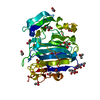


 PDBj
PDBj Assembly
 Homo sapiens neanderthalensis (Neandertal) / References: glutamyl endopeptidase


 Mass: 573.403 Da / Num. of mol.: 2 / Source method: obtained synthetically / Formula: C20H24LuN5O4
 Mass: 59.044 Da / Num. of mol.: 2 / Source method: obtained synthetically / Formula: C2H3O2
 Sample preparation
 / Beamline: XALOC / Wavelength: 1.34 Å
 Processing