 Movie
Movie Controller

[English] 日本語
 Yorodumi
- PDB-7zrq: 1.68 Angstrom crystal structure of Ca/CaM-E140G:CaMKIIdelta pepti... -
+ Open data

- Basic information
| Entry | Database: PDB / ID: 7zrq | |||||||||
|---|---|---|---|---|---|---|---|---|---|---|
| Title | 1.68 Angstrom crystal structure of Ca/CaM-E140G:CaMKIIdelta peptide complex | |||||||||
 Components Components |
| |||||||||
 Keywords Keywords | METAL BINDING PROTEIN / calcium-binding protein / calmodulin / CaM / CaMKII / kinase | |||||||||
| Function / homology |  Function and homology information Function and homology informationregulation of relaxation of cardiac muscle / regulation of cellular localization / negative regulation of sodium ion transmembrane transport / regulation of cardiac muscle cell action potential involved in regulation of contraction / calcium- and calmodulin-dependent protein kinase complex / regulation of cell communication by electrical coupling / Ca2+/calmodulin-dependent protein kinase / regulation of the force of heart contraction / Trafficking of AMPA receptors / endoplasmic reticulum calcium ion homeostasis ...regulation of relaxation of cardiac muscle / regulation of cellular localization / negative regulation of sodium ion transmembrane transport / regulation of cardiac muscle cell action potential involved in regulation of contraction / calcium- and calmodulin-dependent protein kinase complex / regulation of cell communication by electrical coupling / Ca2+/calmodulin-dependent protein kinase / regulation of the force of heart contraction / Trafficking of AMPA receptors / endoplasmic reticulum calcium ion homeostasis / sodium channel inhibitor activity / calcium/calmodulin-dependent protein kinase activity / Assembly and cell surface presentation of NMDA receptors / regulation of calcium ion transmembrane transport via high voltage-gated calcium channel / relaxation of cardiac muscle / CaM pathway / Cam-PDE 1 activation / Sodium/Calcium exchangers / Calmodulin induced events / cardiac muscle cell contraction / Reduction of cytosolic Ca++ levels / Activation of Ca-permeable Kainate Receptor / CREB1 phosphorylation through the activation of CaMKII/CaMKK/CaMKIV cascasde / Loss of phosphorylation of MECP2 at T308 / CREB1 phosphorylation through the activation of Adenylate Cyclase / negative regulation of high voltage-gated calcium channel activity / PKA activation / CaMK IV-mediated phosphorylation of CREB / Glycogen breakdown (glycogenolysis) / negative regulation of ryanodine-sensitive calcium-release channel activity / Activation of RAC1 downstream of NMDARs / organelle localization by membrane tethering / CLEC7A (Dectin-1) induces NFAT activation / : / positive regulation of cardiac muscle hypertrophy / regulation of membrane depolarization / autophagosome membrane docking / negative regulation of calcium ion export across plasma membrane / regulation of ryanodine-sensitive calcium-release channel activity / regulation of cardiac muscle cell action potential / regulation of heart contraction / presynaptic endocytosis / Synthesis of IP3 and IP4 in the cytosol / Phase 0 - rapid depolarisation / Negative regulation of NMDA receptor-mediated neuronal transmission / Unblocking of NMDA receptors, glutamate binding and activation / calcineurin-mediated signaling / RHO GTPases activate PAKs / regulation of heart rate by cardiac conduction / regulation of cell communication by electrical coupling involved in cardiac conduction / Ion transport by P-type ATPases / Uptake and function of anthrax toxins / protein phosphatase activator activity / Long-term potentiation / Calcineurin activates NFAT / HSF1-dependent transactivation / Regulation of MECP2 expression and activity / DARPP-32 events / Smooth Muscle Contraction / regulation of neuronal synaptic plasticity / detection of calcium ion / regulation of cardiac muscle contraction / catalytic complex / regulation of protein localization to plasma membrane / RHO GTPases activate IQGAPs / calcium channel inhibitor activity / presynaptic cytosol / Activation of AMPK downstream of NMDARs / cellular response to interferon-beta / regulation of release of sequestered calcium ion into cytosol by sarcoplasmic reticulum / eNOS activation / Ion homeostasis / Tetrahydrobiopterin (BH4) synthesis, recycling, salvage and regulation / Protein methylation / titin binding / regulation of cardiac muscle contraction by regulation of the release of sequestered calcium ion / regulation of calcium-mediated signaling / voltage-gated potassium channel complex / sarcoplasmic reticulum membrane / positive regulation of cardiac muscle cell apoptotic process / FCERI mediated Ca+2 mobilization / calcium channel complex / substantia nigra development / regulation of heart rate / FCGR3A-mediated IL10 synthesis / cellular response to calcium ion / Antigen activates B Cell Receptor (BCR) leading to generation of second messengers / calyx of Held / Ras activation upon Ca2+ influx through NMDA receptor / adenylate cyclase activator activity / VEGFR2 mediated cell proliferation / VEGFR2 mediated vascular permeability / protein serine/threonine kinase activator activity / regulation of cytokinesis / spindle microtubule / positive regulation of receptor signaling pathway via JAK-STAT / sarcomere / Translocation of SLC2A4 (GLUT4) to the plasma membrane / calcium channel regulator activity / Transcriptional activation of mitochondrial biogenesis Similarity search - Function | |||||||||
| Biological species |  Homo sapiens (human) Homo sapiens (human) | |||||||||
| Method |  X-RAY DIFFRACTION / SYNCHROTRON / MOLECULAR REPLACEMENT / Resolution: 1.68 Å X-RAY DIFFRACTION / SYNCHROTRON / MOLECULAR REPLACEMENT / Resolution: 1.68 Å | |||||||||
 Authors Authors | Helassa, N. / Antonyuk, S. | |||||||||
| Funding support |  United Kingdom, 2items United Kingdom, 2items
| |||||||||
 Citation Citation | Journal: J.Biol.Chem. / Year: 2022 Title: Calmodulin variant E140G associated with long QT syndrome impairs CaMKII delta autophosphorylation and L-type calcium channel inactivation. Authors: Prakash, O. / Gupta, N. / Milburn, A. / McCormick, L. / Deugi, V. / Fisch, P. / Wyles, J. / Thomas, N.L. / Antonyuk, S. / Dart, C. / Helassa, N. | |||||||||
| History |
|
- Structure visualization
| Structure viewer | Molecule: MolmilJmol/JSmol |
|---|
- Downloads & links
-Download
| PDBx/mmCIF format | 7zrq.cif.gz | 79.8 KB | Display | PDBx/mmCIF format |
|---|---|---|---|---|
| PDB format | pdb7zrq.ent.gz | 57.2 KB | Display | PDB format |
| PDBx/mmJSON format | 7zrq.json.gz | Tree view | PDBx/mmJSON format | |
| Others |  Other downloads Other downloads |
-Validation report
| Arichive directory | https://data.pdbj.org/pub/pdb/validation_reports/zr/7zrqftp://data.pdbj.org/pub/pdb/validation_reports/zr/7zrq | HTTPS FTP |
|---|
-Related structure data
| Related structure data | 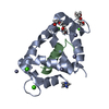 7zrpC 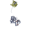 2welS S: Starting model for refinement C: citing same article ( |
|---|---|
| Similar structure data |
-Links
 PDBj
PDBj


- Assembly
| Deposited unit | 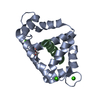
| ||||||||
|---|---|---|---|---|---|---|---|---|---|
| 1 |
| ||||||||
| Unit cell |
|
-Components
| #1: Protein | Mass: 16649.287 Da / Num. of mol.: 1 / Mutation: E140G Source method: isolated from a genetically manipulated source Source: (gene. exp.) Homo sapiens (human) / Gene: CALM1, CALM, CAM, CAM1 / Production host:  | ||||||
|---|---|---|---|---|---|---|---|
| #2: Protein/peptide | Mass: 2515.011 Da / Num. of mol.: 1 / Source method: obtained synthetically / Source: (synth.) Homo sapiens (human)References: UniProt: Q13557, Ca2+/calmodulin-dependent protein kinase | ||||||
| #3: Chemical | ChemComp-CA /   Mass: 40.078 Da / Num. of mol.: 4 / Source method: obtained synthetically / Formula: Ca / Source: (synth.) Homo sapiens (human) / Feature type: SUBJECT OF INVESTIGATION / References: Ca2+/calmodulin-dependent protein kinase Mass: 40.078 Da / Num. of mol.: 4 / Source method: obtained synthetically / Formula: Ca / Source: (synth.) Homo sapiens (human) / Feature type: SUBJECT OF INVESTIGATION / References: Ca2+/calmodulin-dependent protein kinase#4: Chemical | ChemComp-GOL / |   Mass: 92.094 Da / Num. of mol.: 1 / Source method: obtained synthetically / Formula: C3H8O3 Mass: 92.094 Da / Num. of mol.: 1 / Source method: obtained synthetically / Formula: C3H8O3#5: Water | ChemComp-HOH / |  Mass: 18.015 Da / Num. of mol.: 39 / Source method: isolated from a natural source / Formula: H2O Mass: 18.015 Da / Num. of mol.: 39 / Source method: isolated from a natural source / Formula: H2OHas ligand of interest | Y | |
-Experimental details
-Experiment
| Experiment | Method: X-RAY DIFFRACTION / Number of used crystals: 1 |
|---|
- Sample preparation
| Crystal | Density % sol: 27.04 % |
|---|---|
| Crystal grow | Temperature: 292 K / Method: vapor diffusion, hanging drop Details: 0.1 M Na+-HEPES, 0.1 M MOPS (acid), pH 7.5, 0.03 M magnesium chloride hexahydrate, 0.03 M calcium chloride dihydrate, 12.5% v/v MPD; 12.5% PEG 1000; 12.5% w/v PEG 3350 |
-Data collection
| Diffraction | Mean temperature: 100 K / Serial crystal experiment: N |
|---|---|
| Diffraction source | Source: SYNCHROTRON / Site: Diamond / Beamline: I04 / Wavelength: 0.9795 Å |
| Detector | Type: DECTRIS EIGER2 XE 16M / Detector: PIXEL / Date: Dec 11, 2020 |
| Radiation | Protocol: SINGLE WAVELENGTH / Monochromatic (M) / Laue (L): M / Scattering type: x-ray |
| Radiation wavelength | Wavelength: 0.9795 Å / Relative weight: 1 |
| Reflection | Resolution: 1.68→39.59 Å / Num. obs: 15287 / % possible obs: 99.1 % / Redundancy: 6.3 % / CC1/2: 1 / Rmerge(I) obs: 0.048 / Rpim(I) all: 0.03 / Rrim(I) all: 0.057 / Net I/σ(I): 13.5 |
| Reflection shell | Resolution: 1.68→1.71 Å / Redundancy: 4.6 % / Rmerge(I) obs: 2.042 / Mean I/σ(I) obs: 0.6 / Num. unique obs: 684 / CC1/2: 0.311 / Rpim(I) all: 1.452 / Rrim(I) all: 2.522 / % possible all: 87.8 |
- Processing
| Software |
| ||||||||||||||||||||||||||||||||||||||||||||||||||||||||||||||||||||||||||||||||||||||||||||||||||||||||||||||||||||||||||||||||||||||||||||||||||||||||||||||||
|---|---|---|---|---|---|---|---|---|---|---|---|---|---|---|---|---|---|---|---|---|---|---|---|---|---|---|---|---|---|---|---|---|---|---|---|---|---|---|---|---|---|---|---|---|---|---|---|---|---|---|---|---|---|---|---|---|---|---|---|---|---|---|---|---|---|---|---|---|---|---|---|---|---|---|---|---|---|---|---|---|---|---|---|---|---|---|---|---|---|---|---|---|---|---|---|---|---|---|---|---|---|---|---|---|---|---|---|---|---|---|---|---|---|---|---|---|---|---|---|---|---|---|---|---|---|---|---|---|---|---|---|---|---|---|---|---|---|---|---|---|---|---|---|---|---|---|---|---|---|---|---|---|---|---|---|---|---|---|---|---|---|
| Refinement | Method to determine structure: MOLECULAR REPLACEMENT Starting model: 2WEL Resolution: 1.68→39.59 Å / Cor.coef. Fo:Fc: 0.967 / Cor.coef. Fo:Fc free: 0.962 / SU B: 9.797 / SU ML: 0.136 / Cross valid method: THROUGHOUT / ESU R: 0.136 / ESU R Free: 0.128 Details: Hydrogens have been added in their riding positions
| ||||||||||||||||||||||||||||||||||||||||||||||||||||||||||||||||||||||||||||||||||||||||||||||||||||||||||||||||||||||||||||||||||||||||||||||||||||||||||||||||
| Solvent computation | Ion probe radii: 0.8 Å / Shrinkage radii: 0.8 Å / VDW probe radii: 1.2 Å / Solvent model: MASK BULK SOLVENT | ||||||||||||||||||||||||||||||||||||||||||||||||||||||||||||||||||||||||||||||||||||||||||||||||||||||||||||||||||||||||||||||||||||||||||||||||||||||||||||||||
| Displacement parameters | Biso mean: 29.842 Å2
| ||||||||||||||||||||||||||||||||||||||||||||||||||||||||||||||||||||||||||||||||||||||||||||||||||||||||||||||||||||||||||||||||||||||||||||||||||||||||||||||||
| Refinement step | Cycle: LAST / Resolution: 1.68→39.59 Å
| ||||||||||||||||||||||||||||||||||||||||||||||||||||||||||||||||||||||||||||||||||||||||||||||||||||||||||||||||||||||||||||||||||||||||||||||||||||||||||||||||
| Refine LS restraints |
| ||||||||||||||||||||||||||||||||||||||||||||||||||||||||||||||||||||||||||||||||||||||||||||||||||||||||||||||||||||||||||||||||||||||||||||||||||||||||||||||||
| LS refinement shell |
| ||||||||||||||||||||||||||||||||||||||||||||||||||||||||||||||||||||||||||||||||||||||||||||||||||||||||||||||||||||||||||||||||||||||||||||||||||||||||||||||||
| Refinement TLS params. | Method: refined / Refine-ID: X-RAY DIFFRACTION
| ||||||||||||||||||||||||||||||||||||||||||||||||||||||||||||||||||||||||||||||||||||||||||||||||||||||||||||||||||||||||||||||||||||||||||||||||||||||||||||||||
| Refinement TLS group | Selection: ALL |
