 Movie
Movie Controller

[English] 日本語
 Yorodumi
- PDB-7zan: Crystal Structure of human IL-17A in complex with IL-17RA and IL-17RC -
+ Open data

- Basic information
| Entry | Database: PDB / ID: 7zan | ||||||
|---|---|---|---|---|---|---|---|
| Title | Crystal Structure of human IL-17A in complex with IL-17RA and IL-17RC | ||||||
 Components Components |
| ||||||
 Keywords Keywords | CYTOKINE / Immune system | ||||||
| Function / homology |  Function and homology information Function and homology informationinterleukin-17 receptor activity / granulocyte chemotaxis / positive regulation of interleukin-16 production / granulocyte migration / positive regulation of antimicrobial peptide production / Interleukin-17 signaling / T-helper 17 type immune response / cell death / interleukin-17A-mediated signaling pathway / positive regulation of interleukin-23 production ...interleukin-17 receptor activity / granulocyte chemotaxis / positive regulation of interleukin-16 production / granulocyte migration / positive regulation of antimicrobial peptide production / Interleukin-17 signaling / T-helper 17 type immune response / cell death / interleukin-17A-mediated signaling pathway / positive regulation of interleukin-23 production / positive regulation of chemokine (C-X-C motif) ligand 1 production / negative regulation of inflammatory response to wounding / interleukin-17-mediated signaling pathway / fibroblast activation / positive regulation of interleukin-5 production / positive regulation of interleukin-13 production / intestinal epithelial structure maintenance / positive regulation of bicellular tight junction assembly / positive regulation of osteoclast differentiation / positive regulation of cytokine production involved in inflammatory response / mRNA stabilization / keratinocyte proliferation / keratinocyte differentiation / cellular response to interleukin-1 / defense response to fungus / Notch signaling pathway / coreceptor activity / positive regulation of interleukin-12 production / cytokine activity / protein catabolic process / positive regulation of interleukin-1 beta production / response to wounding / positive regulation of interleukin-6 production / response to virus / positive regulation of inflammatory response / positive regulation of tumor necrosis factor production / cell-cell signaling / gene expression / Interleukin-4 and Interleukin-13 signaling / defense response to Gram-negative bacterium / adaptive immune response / cell surface receptor signaling pathway / defense response to Gram-positive bacterium / immune response / inflammatory response / protein heterodimerization activity / external side of plasma membrane / signaling receptor binding / innate immune response / apoptotic process / SARS-CoV-2 activates/modulates innate and adaptive immune responses / cell surface / protein homodimerization activity / positive regulation of transcription by RNA polymerase II / : / extracellular region / plasma membrane Similarity search - Function | ||||||
| Biological species |  Homo sapiens (human) Homo sapiens (human) | ||||||
| Method |  X-RAY DIFFRACTION / SYNCHROTRON / MOLECULAR REPLACEMENT / molecular replacement / Resolution: 5.061 Å X-RAY DIFFRACTION / SYNCHROTRON / MOLECULAR REPLACEMENT / molecular replacement / Resolution: 5.061 Å | ||||||
| Model details | asymmetric unit contains one half complex | ||||||
 Authors Authors | Rondeau, J.M. / Goepfert, A. | ||||||
| Funding support | 1items
| ||||||
 Citation Citation | Journal: Cell Rep / Year: 2022 Title: IL-17-induced dimerization of IL-17RA drives the formation of the IL-17 signalosome to potentiate signaling. Authors: Goepfert, A. / Barske, C. / Lehmann, S. / Wirth, E. / Willemsen, J. / Gudjonsson, J.E. / Ward, N.L. / Sarkar, M.K. / Hemmig, R. / Kolbinger, F. / Rondeau, J.M. | ||||||
| History |
|
- Structure visualization
| Structure viewer | Molecule: MolmilJmol/JSmol |
|---|
- Downloads & links
-Download
| PDBx/mmCIF format | 7zan.cif.gz | 530.5 KB | Display | PDBx/mmCIF format |
|---|---|---|---|---|
| PDB format | pdb7zan.ent.gz | 446.3 KB | Display | PDB format |
| PDBx/mmJSON format | 7zan.json.gz | Tree view | PDBx/mmJSON format | |
| Others |  Other downloads Other downloads |
-Validation report
| Arichive directory | https://data.pdbj.org/pub/pdb/validation_reports/za/7zanftp://data.pdbj.org/pub/pdb/validation_reports/za/7zan | HTTPS FTP |
|---|
-Related structure data
| Related structure data |  5n9bC  4hsaS  6hg4S C: citing same article ( S: Starting model for refinement |
|---|---|
| Similar structure data |
-Links
 PDBj
PDBj

- Assembly
| Deposited unit | 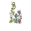
| ||||||||
|---|---|---|---|---|---|---|---|---|---|
| 1 | 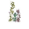
| ||||||||
| Unit cell |
|
-Components
| #1: Protein | Mass: 14177.899 Da / Num. of mol.: 2 / Fragment: IL-17A / Mutation: N68D,C129S Source method: isolated from a genetically manipulated source Source: (gene. exp.) Homo sapiens (human) / Gene: IL17A, CTLA8, IL17 / Plasmid: pCI-derived / Production host: Homo sapiens (human) / Strain (production host): HEK293-6E / References: UniProt: Q16552#2: Protein | | Mass: 34073.570 Da / Num. of mol.: 1 / Mutation: N49D, N206D, N265D Source method: isolated from a genetically manipulated source Source: (gene. exp.) Homo sapiens (human) / Gene: IL17RA, IL17R / Plasmid: pCI-derived / Production host: Homo sapiens (human) / Strain (production host): HEK293S GnTI- / References: UniProt: Q96F46#3: Protein | | Mass: 50558.285 Da / Num. of mol.: 1 / Fragment: Extracellular domain Source method: isolated from a genetically manipulated source Source: (gene. exp.) Homo sapiens (human) / Gene: IL17RC, UNQ6118/PRO20040/PRO38901 / Plasmid: pCI-derived / Production host: Homo sapiens (human) / Strain (production host): HEK293S GnTI- / References: UniProt: Q8NAC3#4: Sugar | ChemComp-NAG /   Type: D-saccharide, beta linking / Mass: 221.208 Da / Num. of mol.: 4 / Source method: obtained synthetically / Formula: C8H15NO6 Type: D-saccharide, beta linking / Mass: 221.208 Da / Num. of mol.: 4 / Source method: obtained synthetically / Formula: C8H15NO6Has ligand of interest | N | Has protein modification | Y | |
|---|
-Experimental details
-Experiment
| Experiment | Method: X-RAY DIFFRACTION / Number of used crystals: 1 |
|---|
- Sample preparation
| Crystal | Density Matthews: 5.28 Å3/Da / Density % sol: 76.72 % / Description: Hexagonal rods |
|---|---|
| Crystal grow | Temperature: 293 K / Method: vapor diffusion, hanging drop / pH: 6.5 / Details: 0.1M MES, 10.0% PEG DME 500 |
-Data collection
| Diffraction | Mean temperature: 100 K / Serial crystal experiment: N | ||||||||||||||||||||||||||||||
|---|---|---|---|---|---|---|---|---|---|---|---|---|---|---|---|---|---|---|---|---|---|---|---|---|---|---|---|---|---|---|---|
| Diffraction source | Source: SYNCHROTRON / Site: SLS  / Beamline: X10SA / Wavelength: 1.00003 Å / Beamline: X10SA / Wavelength: 1.00003 Å | ||||||||||||||||||||||||||||||
| Detector | Type: PSI PILATUS 6M / Detector: PIXEL / Date: Dec 12, 2015 / Details: mirrors | ||||||||||||||||||||||||||||||
| Radiation | Protocol: SINGLE WAVELENGTH / Monochromatic (M) / Laue (L): M / Scattering type: x-ray | ||||||||||||||||||||||||||||||
| Radiation wavelength | Wavelength: 1.00003 Å / Relative weight: 1 | ||||||||||||||||||||||||||||||
| Reflection | Resolution: 5.061→133.601 Å / Num. obs: 10707 / % possible obs: 100 % / Observed criterion σ(I): -3 / Redundancy: 19.2 % / Biso Wilson estimate: 378.5 Å2 / CC1/2: 0.999 / Rmerge(I) obs: 0.103 / Rpim(I) all: 0.025 / Rrim(I) all: 0.106 / Net I/σ(I): 15.8 | ||||||||||||||||||||||||||||||
| Reflection shell | Diffraction-ID: 1
|
-Phasing
| Phasing | Method: molecular replacement |
|---|
- Processing
| Software |
| |||||||||||||||||||||||||||||||||||||||||||||||||||||||||||||||||||||||||||||||||||||||||||||||||||||||||||||||||||||||||||||
|---|---|---|---|---|---|---|---|---|---|---|---|---|---|---|---|---|---|---|---|---|---|---|---|---|---|---|---|---|---|---|---|---|---|---|---|---|---|---|---|---|---|---|---|---|---|---|---|---|---|---|---|---|---|---|---|---|---|---|---|---|---|---|---|---|---|---|---|---|---|---|---|---|---|---|---|---|---|---|---|---|---|---|---|---|---|---|---|---|---|---|---|---|---|---|---|---|---|---|---|---|---|---|---|---|---|---|---|---|---|---|---|---|---|---|---|---|---|---|---|---|---|---|---|---|---|---|
| Refinement | Method to determine structure: MOLECULAR REPLACEMENT Starting model: 4hsa, 6hg4 Resolution: 5.061→37.55 Å / Cor.coef. Fo:Fc: 0.858 / Cor.coef. Fo:Fc free: 0.812 / Cross valid method: THROUGHOUT / σ(F): 0 / SU Rfree Blow DPI: 1.503 Details: HYDROGENS WERE FULLY REFINED WITH ZERO OCCUPANCY AT NUCLEAR POSITION.
| |||||||||||||||||||||||||||||||||||||||||||||||||||||||||||||||||||||||||||||||||||||||||||||||||||||||||||||||||||||||||||||
| Displacement parameters | Biso max: 300 Å2 / Biso mean: 268.1 Å2 / Biso min: 139.46 Å2
| |||||||||||||||||||||||||||||||||||||||||||||||||||||||||||||||||||||||||||||||||||||||||||||||||||||||||||||||||||||||||||||
| Refine analyze | Luzzati coordinate error obs: 0.88 Å | |||||||||||||||||||||||||||||||||||||||||||||||||||||||||||||||||||||||||||||||||||||||||||||||||||||||||||||||||||||||||||||
| Refinement step | Cycle: final / Resolution: 5.061→37.55 Å
| |||||||||||||||||||||||||||||||||||||||||||||||||||||||||||||||||||||||||||||||||||||||||||||||||||||||||||||||||||||||||||||
| Refine LS restraints |
| |||||||||||||||||||||||||||||||||||||||||||||||||||||||||||||||||||||||||||||||||||||||||||||||||||||||||||||||||||||||||||||
| LS refinement shell | Resolution: 5.061→5.15 Å / Rfactor Rfree error: 0
| |||||||||||||||||||||||||||||||||||||||||||||||||||||||||||||||||||||||||||||||||||||||||||||||||||||||||||||||||||||||||||||
| Refinement TLS params. | Method: refined / Refine-ID: X-RAY DIFFRACTION
| |||||||||||||||||||||||||||||||||||||||||||||||||||||||||||||||||||||||||||||||||||||||||||||||||||||||||||||||||||||||||||||
| Refinement TLS group |
|
