 Movie
Movie Controller


+ Open data

- Basic information
| Entry | Database: PDB / ID: 7z5k | ||||||
|---|---|---|---|---|---|---|---|
| Title | Transcription factor MYF5 bound to non-symmetrical site | ||||||
 Components Components |
| ||||||
 Keywords Keywords | TRANSCRIPTION / DNA-binding domain / transcription factor / complex with DNA | ||||||
| Function / homology |  Function and homology information Function and homology informationmuscle cell fate commitment / muscle tissue morphogenesis / positive regulation of skeletal muscle fiber development / embryonic skeletal system morphogenesis / regulation of cell-matrix adhesion / camera-type eye development / TGFBR3 expression / cartilage condensation / muscle organ development / Myogenesis ...muscle cell fate commitment / muscle tissue morphogenesis / positive regulation of skeletal muscle fiber development / embryonic skeletal system morphogenesis / regulation of cell-matrix adhesion / camera-type eye development / TGFBR3 expression / cartilage condensation / muscle organ development / Myogenesis / skeletal muscle cell differentiation / positive regulation of myoblast differentiation / somitogenesis / skeletal muscle tissue development / extracellular matrix organization / ossification / RNA polymerase II transcription regulator complex / DNA-binding transcription activator activity, RNA polymerase II-specific / DNA-binding transcription factor activity, RNA polymerase II-specific / protein dimerization activity / RNA polymerase II cis-regulatory region sequence-specific DNA binding / regulation of transcription by RNA polymerase II / chromatin / nucleoplasm Similarity search - Function | ||||||
| Biological species |  Homo sapiens (human) Homo sapiens (human) | ||||||
| Method |  X-RAY DIFFRACTION / SYNCHROTRON / MOLECULAR REPLACEMENT / molecular replacement / Resolution: 2.28 Å X-RAY DIFFRACTION / SYNCHROTRON / MOLECULAR REPLACEMENT / molecular replacement / Resolution: 2.28 Å | ||||||
 Authors Authors | Morgunova, E. / Popov, A. / Yin, Y. / Taipale, J. | ||||||
| Funding support | 1items
| ||||||
 Citation Citation | Journal: Nat.Struct.Mol.Biol. / Year: 2025 Title: Interfacial water confers transcription factors with dinucleotide specificity. Authors: Morgunova, E. / Nagy, G. / Yin, Y. / Zhu, F. / Nayak, S.P. / Xiao, T. / Sokolov, I. / Popov, A. / Laughton, C. / Grubmuller, H. / Taipale, J. | ||||||
| History |
|
- Structure visualization
| Structure viewer | Molecule: MolmilJmol/JSmol |
|---|
- Downloads & links
-Download
| PDBx/mmCIF format | 7z5k.cif.gz | 60.6 KB | Display | PDBx/mmCIF format |
|---|---|---|---|---|
| PDB format | pdb7z5k.ent.gz | 40.4 KB | Display | PDB format |
| PDBx/mmJSON format | 7z5k.json.gz | Tree view | PDBx/mmJSON format | |
| Others |  Other downloads Other downloads |
-Validation report
| Arichive directory | https://data.pdbj.org/pub/pdb/validation_reports/z5/7z5kftp://data.pdbj.org/pub/pdb/validation_reports/z5/7z5k | HTTPS FTP |
|---|
-Related structure data
| Related structure data | 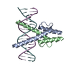 7z5iC 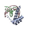 8pm5C 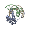 8pm7C 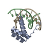 8pmcC 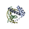 8pmfC 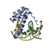 8pmnC 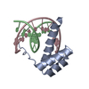 8pmvC 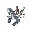 8pn4C 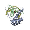 8pnaC 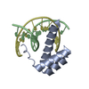 8pncC 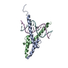 1mdyS S: Starting model for refinement C: citing same article ( |
|---|---|
| Similar structure data |
-Links
 PDBj
PDBj


- Assembly
| Deposited unit | 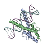
| ||||||||
|---|---|---|---|---|---|---|---|---|---|
| 1 |
| ||||||||
| Unit cell |
|
-Components
| #1: Protein | Mass: 6888.085 Da / Num. of mol.: 2 Source method: isolated from a genetically manipulated source Source: (gene. exp.) Homo sapiens (human) / Gene: MYF5, BHLHC2 / Production host:  #2: DNA chain | | Mass: 5526.581 Da / Num. of mol.: 1 / Source method: obtained synthetically / Source: (synth.) Homo sapiens (human)#3: DNA chain | | Mass: 5508.553 Da / Num. of mol.: 1 / Source method: obtained synthetically / Source: (synth.) Homo sapiens (human)#4: Water | ChemComp-HOH / |  Mass: 18.015 Da / Num. of mol.: 76 / Source method: isolated from a natural source / Formula: H2O Mass: 18.015 Da / Num. of mol.: 76 / Source method: isolated from a natural source / Formula: H2OHas protein modification | N | |
|---|
-Experimental details
-Experiment
| Experiment | Method: X-RAY DIFFRACTION / Number of used crystals: 1 |
|---|
- Sample preparation
| Crystal | Density Matthews: 3.13 Å3/Da / Density % sol: 60.72 % |
|---|---|
| Crystal grow | Temperature: 295 K / Method: vapor diffusion, hanging drop / pH: 4.5 Details: 13% PEG 1000, 2.5% MPD, 6,25% PEG 400, 0.05M Sodium Acetate, pH 4.5 |
-Data collection
| Diffraction | Mean temperature: 100 K / Serial crystal experiment: N | ||||||||||||||||||||||||||||||
|---|---|---|---|---|---|---|---|---|---|---|---|---|---|---|---|---|---|---|---|---|---|---|---|---|---|---|---|---|---|---|---|
| Diffraction source | Source: SYNCHROTRON / Site: ESRF  / Beamline: ID23-1 / Wavelength: 0.9724 Å / Beamline: ID23-1 / Wavelength: 0.9724 Å | ||||||||||||||||||||||||||||||
| Detector | Type: DECTRIS PILATUS 6M / Detector: PIXEL / Date: Aug 8, 2018 | ||||||||||||||||||||||||||||||
| Radiation | Protocol: SINGLE WAVELENGTH / Monochromatic (M) / Laue (L): M / Scattering type: x-ray | ||||||||||||||||||||||||||||||
| Radiation wavelength | Wavelength: 0.9724 Å / Relative weight: 1 | ||||||||||||||||||||||||||||||
| Reflection | Resolution: 1.99→46 Å / Num. obs: 26259 / % possible obs: 93.1 % / Redundancy: 4.6 % / Biso Wilson estimate: 57.98 Å2 / CC1/2: 0.996 / Rmerge(I) obs: 0.137 / Rpim(I) all: 0.069 / Rrim(I) all: 0.154 / Net I/σ(I): 3.4 | ||||||||||||||||||||||||||||||
| Reflection shell | Diffraction-ID: 1
|
-Phasing
| Phasing | Method: molecular replacement |
|---|
- Processing
| Software |
| ||||||||||||||||||||||||||||||||||||||||||||||||||||||||||||
|---|---|---|---|---|---|---|---|---|---|---|---|---|---|---|---|---|---|---|---|---|---|---|---|---|---|---|---|---|---|---|---|---|---|---|---|---|---|---|---|---|---|---|---|---|---|---|---|---|---|---|---|---|---|---|---|---|---|---|---|---|---|
| Refinement | Method to determine structure: MOLECULAR REPLACEMENT Starting model: 1MDY Resolution: 2.28→45.998 Å / SU ML: 0.39 / Cross valid method: THROUGHOUT / σ(F): 0.04 / Phase error: 35.63 / Stereochemistry target values: ML
| ||||||||||||||||||||||||||||||||||||||||||||||||||||||||||||
| Solvent computation | Shrinkage radii: 0.9 Å / VDW probe radii: 1.11 Å / Solvent model: FLAT BULK SOLVENT MODEL | ||||||||||||||||||||||||||||||||||||||||||||||||||||||||||||
| Displacement parameters | Biso max: 176.93 Å2 / Biso mean: 84.145 Å2 / Biso min: 30 Å2 | ||||||||||||||||||||||||||||||||||||||||||||||||||||||||||||
| Refinement step | Cycle: final / Resolution: 2.28→45.998 Å
| ||||||||||||||||||||||||||||||||||||||||||||||||||||||||||||
| LS refinement shell | Refine-ID: X-RAY DIFFRACTION / Rfactor Rfree error: 0
|
