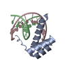
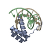
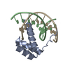
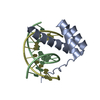
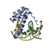
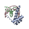
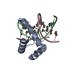
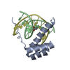
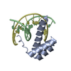
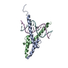
登録情報 データベース : PDB / ID : 7z5iタイトル Transcription factor MYF5 bound to symmetrical site DNA (5'-D(P*AP*CP*GP*CP*GP*TP*CP*AP*GP*CP*TP*GP*AP*CP*GP*CP*GP*C)-3')DNA (5'-D(P*GP*CP*GP*CP*GP*TP*CP*AP*GP*CP*TP*GP*AP*CP*GP*CP*GP*T)-3')Myogenic factor 5 キーワード / / / 機能・相同性 分子機能 ドメイン・相同性 構成要素
/ / / / / / / / / / / / / / / / / / / / / / / / / / / / / / / / / / / / / / / / / / 生物種 Homo sapiens (ヒト)手法 / / / 解像度 : 3 Å データ登録者 Morgunova, E. / Popov, A. / Yin, Y. / Taipale, J. 資金援助 1件 ジャーナル : Nat.Struct.Mol.Biol. / 年 : 2025タイトル : Interfacial water confers transcription factors with dinucleotide specificity.著者 : Morgunova, E. / Nagy, G. / Yin, Y. / Zhu, F. / Nayak, S.P. / Xiao, T. / Sokolov, I. / Popov, A. / Laughton, C. / Grubmuller, H. / Taipale, J. 履歴 登録 2022年3月9日 登録サイト / 処理サイト 改定 1.0 2023年3月22日 Provider / タイプ 改定 1.1 2024年2月7日 Group / Refinement descriptionカテゴリ chem_comp_atom / chem_comp_bond ... chem_comp_atom / chem_comp_bond / pdbx_initial_refinement_model / struct_ncs_dom_lim Item _struct_ncs_dom_lim.beg_auth_comp_id / _struct_ncs_dom_lim.beg_label_asym_id ... _struct_ncs_dom_lim.beg_auth_comp_id / _struct_ncs_dom_lim.beg_label_asym_id / _struct_ncs_dom_lim.beg_label_comp_id / _struct_ncs_dom_lim.beg_label_seq_id / _struct_ncs_dom_lim.end_auth_comp_id / _struct_ncs_dom_lim.end_label_asym_id / _struct_ncs_dom_lim.end_label_comp_id / _struct_ncs_dom_lim.end_label_seq_id 改定 1.2 2025年1月15日 Group / Structure summaryカテゴリ / citation_author / pdbx_entry_detailsItem _citation.country / _citation.journal_abbrev ... _citation.country / _citation.journal_abbrev / _citation.journal_id_CSD / _citation.journal_id_ISSN / _citation.pdbx_database_id_DOI / _citation.pdbx_database_id_PubMed / _citation.title / _citation.year
すべて表示 表示を減らす
 ムービー
ムービー コントローラー


 データを開く
 基本情報
基本情報 要素
要素 キーワード
キーワード 機能・相同性情報
 Homo sapiens (ヒト)
 X線回折 /
  データ登録者
 引用
引用 構造の表示
構造の表示 ダウンロードとリンク
 その他のダウンロード
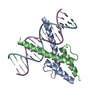
 PDBj
PDBj


 集合体
集合体

 分子量: 18.015 Da / 分子数: 3 / 由来タイプ: 天然 / 式: H2O
 試料調製
試料調製 / ビームライン: ID23-1 / 波長: 0.972 Å
 解析
解析