 Movie
Movie Controller


+ Open data

- Basic information
| Entry | Database: PDB / ID: 7z1p | ||||||
|---|---|---|---|---|---|---|---|
| Title | X-ray crystal structure of SLPYL1-E151D mutant | ||||||
 Components Components | SLPYL1-E151D | ||||||
 Keywords Keywords | PLANT PROTEIN / SLPYL1 / ABA receptor protein | ||||||
| Function / homology |  Function and homology information Function and homology informationprotein phosphatase inhibitor complex / abscisic acid binding / abscisic acid-activated signaling pathway / protein phosphatase inhibitor activity / signaling receptor activity / protein homodimerization activity / nucleus / cytoplasm Similarity search - Function | ||||||
| Biological species |  | ||||||
| Method |  X-RAY DIFFRACTION / SYNCHROTRON / MOLECULAR REPLACEMENT / Resolution: 1.82 Å X-RAY DIFFRACTION / SYNCHROTRON / MOLECULAR REPLACEMENT / Resolution: 1.82 Å | ||||||
 Authors Authors | Infantes, L. / Albert, A. | ||||||
| Funding support |  Spain, 1items Spain, 1items
| ||||||
 Citation Citation | Journal: Front Plant Sci / Year: 2022 Title: Structure-Based Modulation of the Ligand Sensitivity of a Tomato Dimeric Abscisic Acid Receptor Through a Glu to Asp Mutation in the Latch Loop. Authors: Infantes, L. / Rivera-Moreno, M. / Daniel-Mozo, M. / Benavente, J.L. / Ocana-Cuesta, J. / Coego, A. / Lozano-Juste, J. / Rodriguez, P.L. / Albert, A. | ||||||
| History |
|
- Structure visualization
| Structure viewer | Molecule: MolmilJmol/JSmol |
|---|
- Downloads & links
-Download
| PDBx/mmCIF format | 7z1p.cif.gz | 109.9 KB | Display | PDBx/mmCIF format |
|---|---|---|---|---|
| PDB format | pdb7z1p.ent.gz | 70 KB | Display | PDB format |
| PDBx/mmJSON format | 7z1p.json.gz | Tree view | PDBx/mmJSON format | |
| Others |  Other downloads Other downloads |
-Validation report
| Arichive directory | https://data.pdbj.org/pub/pdb/validation_reports/z1/7z1pftp://data.pdbj.org/pub/pdb/validation_reports/z1/7z1p | HTTPS FTP |
|---|
-Related structure data
| Related structure data | 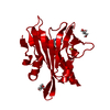 7z1qC  7z1rC  7z1sC  5moaS S: Starting model for refinement C: citing same article ( |
|---|---|
| Similar structure data |

-Links
 PDBj
PDBj- Assembly
| Deposited unit | 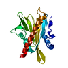
| ||||||||||||
|---|---|---|---|---|---|---|---|---|---|---|---|---|---|
| 1 | 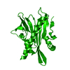
| ||||||||||||
| Unit cell |
|
-Components
| #1: Protein | Mass: 25724.418 Da / Num. of mol.: 1 / Mutation: E151D Source method: isolated from a genetically manipulated source Source: (gene. exp.)  |
|---|---|
| #2: Water | ChemComp-HOH /  Mass: 18.015 Da / Num. of mol.: 72 / Source method: isolated from a natural source / Formula: H2O Mass: 18.015 Da / Num. of mol.: 72 / Source method: isolated from a natural source / Formula: H2O |
-Experimental details
-Experiment
| Experiment | Method: X-RAY DIFFRACTION / Number of used crystals: 1 |
|---|
- Sample preparation
| Crystal | Density Matthews: 2.35 Å3/Da / Density % sol: 47.69 % |
|---|---|
| Crystal grow | Temperature: 291 K / Method: vapor diffusion, hanging drop / pH: 7 / Details: Ammonium sulfate 1.8M |
-Data collection
| Diffraction | Mean temperature: 100 K / Serial crystal experiment: N |
|---|---|
| Diffraction source | Source: SYNCHROTRON / Site: ALBA / Beamline: XALOC / Wavelength: 0.979261 Å |
| Detector | Type: DECTRIS PILATUS 6M / Detector: PIXEL / Date: May 5, 2021 |
| Radiation | Protocol: SINGLE WAVELENGTH / Monochromatic (M) / Laue (L): M / Scattering type: x-ray |
| Radiation wavelength | Wavelength: 0.979261 Å / Relative weight: 1 |
| Reflection | Resolution: 1.82→44.98 Å / Num. obs: 21962 / % possible obs: 99.76 % / Redundancy: 16.4 % / Biso Wilson estimate: 38.73 Å2 / CC1/2: 1 / CC star: 1 / Rmerge(I) obs: 0.05691 / Rpim(I) all: 0.01465 / Rrim(I) all: 0.05881 / Net I/σ(I): 23.84 |
| Reflection shell | Resolution: 1.82→1.885 Å / Redundancy: 16.3 % / Rmerge(I) obs: 1.979 / Mean I/σ(I) obs: 1.77 / Num. unique obs: 2149 / CC1/2: 0.896 / Rpim(I) all: 0.4977 / Rrim(I) all: 2.042 / % possible all: 99.16 |
- Processing
| Software |
| |||||||||||||||||||||||||||||||||||||||||||||||||||||||||||||||
|---|---|---|---|---|---|---|---|---|---|---|---|---|---|---|---|---|---|---|---|---|---|---|---|---|---|---|---|---|---|---|---|---|---|---|---|---|---|---|---|---|---|---|---|---|---|---|---|---|---|---|---|---|---|---|---|---|---|---|---|---|---|---|---|---|
| Refinement | Method to determine structure: MOLECULAR REPLACEMENT Starting model: 5MOA Resolution: 1.82→44.98 Å / SU ML: 0.2869 / Cross valid method: FREE R-VALUE / σ(F): 1.34 / Phase error: 38.1665 Stereochemistry target values: GeoStd + Monomer Library + CDL v1.2
| |||||||||||||||||||||||||||||||||||||||||||||||||||||||||||||||
| Solvent computation | Shrinkage radii: 0.9 Å / VDW probe radii: 1.1 Å / Solvent model: FLAT BULK SOLVENT MODEL | |||||||||||||||||||||||||||||||||||||||||||||||||||||||||||||||
| Displacement parameters | Biso mean: 52.06 Å2 | |||||||||||||||||||||||||||||||||||||||||||||||||||||||||||||||
| Refinement step | Cycle: LAST / Resolution: 1.82→44.98 Å
| |||||||||||||||||||||||||||||||||||||||||||||||||||||||||||||||
| Refine LS restraints |
| |||||||||||||||||||||||||||||||||||||||||||||||||||||||||||||||
| LS refinement shell |
| |||||||||||||||||||||||||||||||||||||||||||||||||||||||||||||||
| Refinement TLS params. | Method: refined / Origin x: 1.63014664679 Å / Origin y: 30.8149453295 Å / Origin z: -5.41587322678 Å
| |||||||||||||||||||||||||||||||||||||||||||||||||||||||||||||||
| Refinement TLS group | Selection details: all |
