 Movie
Movie Controller


+ Open data

- Basic information
| Entry | Database: PDB / ID: 5mob | ||||||
|---|---|---|---|---|---|---|---|
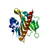
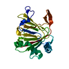
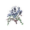
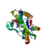
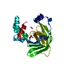
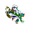
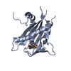
| Title | ABA RECEPTOR FROM TOMATO, SlPYL1 | ||||||
 Components Components | SlPYL1_ABA | ||||||
 Keywords Keywords | SIGNALING PROTEIN / ABA / RECEPTOR / SIGNALING / STRESS | ||||||
| Function / homology | START domain / Alpha-D-Glucose-1,6-Bisphosphate; Chain A, domain 4 / 2-Layer Sandwich / Alpha Beta / Chem-A8S / :  Function and homology information Function and homology information | ||||||
| Biological species |  | ||||||
| Method |  X-RAY DIFFRACTION / SYNCHROTRON / MOLECULAR REPLACEMENT / Resolution: 1.669 Å X-RAY DIFFRACTION / SYNCHROTRON / MOLECULAR REPLACEMENT / Resolution: 1.669 Å | ||||||
 Authors Authors | Moreno-Alvero, M. / Yunta, C. / Gonzalez-Guzman, M. / Arbona, V. / Granell, A. / Martinez-Ripoll, M. / Infantes, L. / Rodriguez, P.L. / Albert, A. | ||||||
 Citation Citation | Journal: Mol Plant / Year: 2017 Title: Structure of Ligand-Bound Intermediates of Crop ABA Receptors Highlights PP2C as Necessary ABA Co-receptor. Authors: Moreno-Alvero, M. / Yunta, C. / Gonzalez-Guzman, M. / Lozano-Juste, J. / Benavente, J.L. / Arbona, V. / Menendez, M. / Martinez-Ripoll, M. / Infantes, L. / Gomez-Cadenas, A. / Rodriguez, P.L. / Albert, A. | ||||||
| History |
|
- Structure visualization
| Structure viewer | Molecule: MolmilJmol/JSmol |
|---|
- Downloads & links
-Download
| PDBx/mmCIF format | 5mob.cif.gz | 94.7 KB | Display | PDBx/mmCIF format |
|---|---|---|---|---|
| PDB format | pdb5mob.ent.gz | 71.1 KB | Display | PDB format |
| PDBx/mmJSON format | 5mob.json.gz | Tree view | PDBx/mmJSON format | |
| Others |  Other downloads Other downloads |
-Validation report
| Arichive directory | https://data.pdbj.org/pub/pdb/validation_reports/mo/5mobftp://data.pdbj.org/pub/pdb/validation_reports/mo/5mob | HTTPS FTP |
|---|
-Related structure data


-Links
 PDBj
PDBj- Assembly
| Deposited unit | 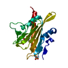
| ||||||||
|---|---|---|---|---|---|---|---|---|---|
| 1 |
| ||||||||
| Unit cell |
|
-Components
| #1: Protein | Mass: 25738.443 Da / Num. of mol.: 1 Source method: isolated from a genetically manipulated source Source: (gene. exp.)  |
|---|---|
| #2: Chemical | ChemComp-A8S / (  Mass: 264.317 Da / Num. of mol.: 1 / Source method: obtained synthetically / Formula: C15H20O4 / Comment: hormone*YM Mass: 264.317 Da / Num. of mol.: 1 / Source method: obtained synthetically / Formula: C15H20O4 / Comment: hormone*YM |
| #3: Chemical | ChemComp-SO4 /   Mass: 96.063 Da / Num. of mol.: 1 / Source method: obtained synthetically / Formula: SO4 Mass: 96.063 Da / Num. of mol.: 1 / Source method: obtained synthetically / Formula: SO4 |
| #4: Water | ChemComp-HOH /  Mass: 18.015 Da / Num. of mol.: 168 / Source method: isolated from a natural source / Formula: H2O Mass: 18.015 Da / Num. of mol.: 168 / Source method: isolated from a natural source / Formula: H2O |
-Experimental details
-Experiment
| Experiment | Method: X-RAY DIFFRACTION / Number of used crystals: 1 |
|---|
- Sample preparation
| Crystal | Density Matthews: 2.46 Å3/Da / Density % sol: 50.08 % |
|---|---|
| Crystal grow | Temperature: 295 K / Method: microbatch / pH: 7 / Details: 3.5 M de sulfato de amonio pH 7.0 |
-Data collection
| Diffraction | Mean temperature: 100 K |
|---|---|
| Diffraction source | Source: SYNCHROTRON / Site: ESRF  / Beamline: ID23-1 / Wavelength: 0.93 Å / Beamline: ID23-1 / Wavelength: 0.93 Å |
| Detector | Type: ADSC QUANTUM 210 / Detector: CCD / Date: Apr 25, 2014 |
| Radiation | Protocol: SINGLE WAVELENGTH / Monochromatic (M) / Laue (L): M / Scattering type: x-ray |
| Radiation wavelength | Wavelength: 0.93 Å / Relative weight: 1 |
| Reflection | Resolution: 1.67→28.63 Å / Num. obs: 27365 / % possible obs: 99.9 % / Redundancy: 6.1 % / CC1/2: 0.99 / Net I/σ(I): 17.1 |
| Reflection shell | Resolution: 1.67→1.7 Å / Redundancy: 6.1 % / % possible all: 100 |
- Processing
| Software |
| |||||||||||||||||||||||||||||||||||||||||||||||||||||||||||||||||||||||||||||||||||||||||||||||||||||||||||||||||||||||||||||||||||||||||||||||||||
|---|---|---|---|---|---|---|---|---|---|---|---|---|---|---|---|---|---|---|---|---|---|---|---|---|---|---|---|---|---|---|---|---|---|---|---|---|---|---|---|---|---|---|---|---|---|---|---|---|---|---|---|---|---|---|---|---|---|---|---|---|---|---|---|---|---|---|---|---|---|---|---|---|---|---|---|---|---|---|---|---|---|---|---|---|---|---|---|---|---|---|---|---|---|---|---|---|---|---|---|---|---|---|---|---|---|---|---|---|---|---|---|---|---|---|---|---|---|---|---|---|---|---|---|---|---|---|---|---|---|---|---|---|---|---|---|---|---|---|---|---|---|---|---|---|---|---|---|---|
| Refinement | Method to determine structure: MOLECULAR REPLACEMENT / Resolution: 1.669→28.631 Å / SU ML: 0.41 / Cross valid method: NONE / σ(F): 0.69 / Phase error: 27.33 / Stereochemistry target values: ML
| |||||||||||||||||||||||||||||||||||||||||||||||||||||||||||||||||||||||||||||||||||||||||||||||||||||||||||||||||||||||||||||||||||||||||||||||||||
| Solvent computation | Shrinkage radii: 0.86 Å / VDW probe radii: 1.1 Å / Solvent model: FLAT BULK SOLVENT MODEL / Bsol: 62.757 Å2 / ksol: 0.4 e/Å3 | |||||||||||||||||||||||||||||||||||||||||||||||||||||||||||||||||||||||||||||||||||||||||||||||||||||||||||||||||||||||||||||||||||||||||||||||||||
| Displacement parameters |
| |||||||||||||||||||||||||||||||||||||||||||||||||||||||||||||||||||||||||||||||||||||||||||||||||||||||||||||||||||||||||||||||||||||||||||||||||||
| Refinement step | Cycle: LAST / Resolution: 1.669→28.631 Å
| |||||||||||||||||||||||||||||||||||||||||||||||||||||||||||||||||||||||||||||||||||||||||||||||||||||||||||||||||||||||||||||||||||||||||||||||||||
| Refine LS restraints |
| |||||||||||||||||||||||||||||||||||||||||||||||||||||||||||||||||||||||||||||||||||||||||||||||||||||||||||||||||||||||||||||||||||||||||||||||||||
| LS refinement shell |
| |||||||||||||||||||||||||||||||||||||||||||||||||||||||||||||||||||||||||||||||||||||||||||||||||||||||||||||||||||||||||||||||||||||||||||||||||||
| Refinement TLS params. | Method: refined / Origin x: 25.5726 Å / Origin y: 15.1959 Å / Origin z: 3.8631 Å
| |||||||||||||||||||||||||||||||||||||||||||||||||||||||||||||||||||||||||||||||||||||||||||||||||||||||||||||||||||||||||||||||||||||||||||||||||||
| Refinement TLS group | Selection details: all |
