 Movie
Movie Controller

[English] 日本語
 Yorodumi
- PDB-7ykb: Neutron Structure of PcyA D105N Mutant Complexed with Biliverdin ... -
+ Open data

- Basic information
| Entry | Database: PDB / ID: 7ykb | ||||||
|---|---|---|---|---|---|---|---|
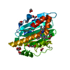
| Title | Neutron Structure of PcyA D105N Mutant Complexed with Biliverdin at Room Temperature | ||||||
 Components Components | Phycocyanobilin:ferredoxin oxidoreductase | ||||||
 Keywords Keywords | OXIDOREDUCTASE / phycocyanobilin / ferredoxin oxi-doreductase / neutron / hydrogen / proton / mutant / reaction / absorption spectrum | ||||||
| Function / homology | phycocyanobilin:ferredoxin oxidoreductase / phycocyanobilin:ferredoxin oxidoreductase activity / Phycocyanobilin:ferredoxin oxidoreductase / Ferredoxin-dependent bilin reductase / Ferredoxin-dependent bilin reductase / phytochromobilin biosynthetic process / cobalt ion binding / Chem-BLR / Phycocyanobilin:ferredoxin oxidoreductase Function and homology information Function and homology information | ||||||
| Biological species |  | ||||||
| Method |  X-RAY DIFFRACTION / NEUTRON DIFFRACTION / SYNCHROTRON / MOLECULAR REPLACEMENT / Resolution: 1.38 Å X-RAY DIFFRACTION / NEUTRON DIFFRACTION / SYNCHROTRON / MOLECULAR REPLACEMENT / Resolution: 1.38 Å | ||||||
 Authors Authors | Unno, M. / Nanasawa, R. | ||||||
| Funding support |  Japan, 1items Japan, 1items
| ||||||
 Citation Citation | Journal: J.Biol.Chem. / Year: 2022 Title: Neutron crystallography and quantum chemical analysis of bilin reductase PcyA mutants reveal substrate and catalytic residue protonation states. Authors: Joutsuka, T. / Nanasawa, R. / Igarashi, K. / Horie, K. / Sugishima, M. / Hagiwara, Y. / Wada, K. / Fukuyama, K. / Yano, N. / Mori, S. / Ostermann, A. / Kusaka, K. / Unno, M. | ||||||
| History |
|
- Structure visualization
| Structure viewer | Molecule: MolmilJmol/JSmol |
|---|
- Downloads & links
-Download
| PDBx/mmCIF format | 7ykb.cif.gz | 161.6 KB | Display | PDBx/mmCIF format |
|---|---|---|---|---|
| PDB format | pdb7ykb.ent.gz | 107.7 KB | Display | PDB format |
| PDBx/mmJSON format | 7ykb.json.gz | Tree view | PDBx/mmJSON format | |
| Others |  Other downloads Other downloads |
-Validation report
| Arichive directory | https://data.pdbj.org/pub/pdb/validation_reports/yk/7ykbftp://data.pdbj.org/pub/pdb/validation_reports/yk/7ykb | HTTPS FTP |
|---|
-Related structure data
| Related structure data |  7yk9C  3f0mS C: citing same article ( S: Starting model for refinement |
|---|---|
| Similar structure data |

-Links
 PDBj
PDBj- Assembly
| Deposited unit | 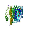
| ||||||||||||
|---|---|---|---|---|---|---|---|---|---|---|---|---|---|
| 1 |
| ||||||||||||
| Unit cell |
| ||||||||||||
| Components on special symmetry positions |
|
-Components
| #1: Protein | Mass: 28155.172 Da / Num. of mol.: 1 / Mutation: D105N Source method: isolated from a genetically manipulated source Source: (gene. exp.) Strain: Kazusa / Gene: pcyA / Production host: References: UniProt: Q55891, phycocyanobilin:ferredoxin oxidoreductase |
|---|---|
| #2: Chemical | ChemComp-BLR /   Mass: 584.662 Da / Num. of mol.: 1 / Source method: obtained synthetically / Formula: C33H36N4O6 / Feature type: SUBJECT OF INVESTIGATION Mass: 584.662 Da / Num. of mol.: 1 / Source method: obtained synthetically / Formula: C33H36N4O6 / Feature type: SUBJECT OF INVESTIGATION |
| #3: Chemical | ChemComp-NA /   Mass: 22.990 Da / Num. of mol.: 1 / Source method: obtained synthetically / Formula: Na Mass: 22.990 Da / Num. of mol.: 1 / Source method: obtained synthetically / Formula: Na |
| #4: Water | ChemComp-HOH /  Mass: 18.015 Da / Num. of mol.: 219 / Source method: isolated from a natural source / Formula: H2O Mass: 18.015 Da / Num. of mol.: 219 / Source method: isolated from a natural source / Formula: H2O |
| Has ligand of interest | Y |
-Experimental details
-Experiment
| Experiment |
|
|---|
- Sample preparation
| Crystal | Density Matthews: 2.74 Å3/Da / Density % sol: 55.13 % |
|---|---|
| Crystal grow | Temperature: 293 K / Method: vapor diffusion, sitting drop / Details: NaCl, ammonium sulfate, MES |
-Data collection
| Diffraction |
| |||||||||||||||||||||
|---|---|---|---|---|---|---|---|---|---|---|---|---|---|---|---|---|---|---|---|---|---|---|
| Diffraction source |
| |||||||||||||||||||||
| Detector |
| |||||||||||||||||||||
| Radiation |
| |||||||||||||||||||||
| Radiation wavelength |
| |||||||||||||||||||||
| Reflection | Biso Wilson estimate: 15.07 Å2 / Entry-ID: 7YKB
| |||||||||||||||||||||
| Reflection shell |
|
- Processing
| Software |
| |||||||||||||||||||||||||||||||||||||||||||||||||||||||||||||||
|---|---|---|---|---|---|---|---|---|---|---|---|---|---|---|---|---|---|---|---|---|---|---|---|---|---|---|---|---|---|---|---|---|---|---|---|---|---|---|---|---|---|---|---|---|---|---|---|---|---|---|---|---|---|---|---|---|---|---|---|---|---|---|---|---|
| Refinement | SU ML: 0.1322 / Cross valid method: FREE R-VALUE / Method to determine structure:
| |||||||||||||||||||||||||||||||||||||||||||||||||||||||||||||||
| Refinement step | Cycle: LAST / Resolution: 1.38→34.58 Å
| |||||||||||||||||||||||||||||||||||||||||||||||||||||||||||||||
| Refine LS restraints |
| |||||||||||||||||||||||||||||||||||||||||||||||||||||||||||||||
| LS refinement shell |
|
