 Movie
Movie Controller


+ Open data

- Basic information
| Entry | Database: PDB / ID: 7y5r | ||||||
|---|---|---|---|---|---|---|---|
| Title | Crystal structure of sDscam FNIII2 domain, isoform alpha7 | ||||||
 Components Components | Down Syndrome Cell Adhesion Molecules | ||||||
 Keywords Keywords | CELL ADHESION / cell surface receptor | ||||||
| Biological species |  Chelicerata (chelicerates) Chelicerata (chelicerates) | ||||||
| Method |  X-RAY DIFFRACTION / SYNCHROTRON / MOLECULAR REPLACEMENT / Resolution: 1.562 Å X-RAY DIFFRACTION / SYNCHROTRON / MOLECULAR REPLACEMENT / Resolution: 1.562 Å | ||||||
 Authors Authors | Chen, Q. / Yu, Y. / Cheng, J. | ||||||
| Funding support |  China, 1items China, 1items
| ||||||
 Citation Citation | Journal: Nat Commun / Year: 2023 Title: Structural basis for the self-recognition of sDSCAM in Chelicerata. Authors: Cheng, J. / Yu, Y. / Wang, X. / Zheng, X. / Liu, T. / Hu, D. / Jin, Y. / Lai, Y. / Fu, T.M. / Chen, Q. | ||||||
| History |
|
- Structure visualization
| Structure viewer | Molecule:  MolmilJmol/JSmol MolmilJmol/JSmol |
|---|
- Downloads & links
-Download
| PDBx/mmCIF format | 7y5r.cif.gz | 55.8 KB | Display | PDBx/mmCIF format |
|---|---|---|---|---|
| PDB format | pdb7y5r.ent.gz | 39.2 KB | Display | PDB format |
| PDBx/mmJSON format | 7y5r.json.gz | Tree view | PDBx/mmJSON format | |
| Others |  Other downloads Other downloads |
-Validation report
| Summary document | 7y5r_validation.pdf.gz | 1.2 MB | Display | wwPDB validaton report |
|---|---|---|---|---|
| Full document | 7y5r_full_validation.pdf.gz | 1.2 MB | Display | |
| Data in XML | 7y5r_validation.xml.gz | 8.3 KB | Display | |
| Data in CIF | 7y5r_validation.cif.gz | 11.5 KB | Display | |
| Arichive directory | https://data.pdbj.org/pub/pdb/validation_reports/y5/7y5rftp://data.pdbj.org/pub/pdb/validation_reports/y5/7y5r | HTTPS FTP |
-Related structure data
| Related structure data | 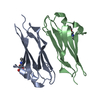 7y4xC 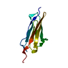 7y54C 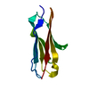 7y5jC  7y6eC 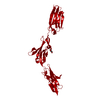 7y6oC 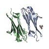 7y73C  7y8hC  7y8iC  7y8sC  7y95C 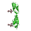 7y9aC  1va9S S: Starting model for refinement C: citing same article ( |
|---|
-Links
 PDBj
PDBj- Assembly
| Deposited unit | 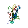
| ||||||||
|---|---|---|---|---|---|---|---|---|---|
| 1 |
| ||||||||
| Unit cell |
|
-Components
| #1: Protein | Mass: 11477.840 Da / Num. of mol.: 1 Source method: isolated from a genetically manipulated source Source: (gene. exp.) Chelicerata (chelicerates) / Production host:  | ||||
|---|---|---|---|---|---|
| #2: Chemical |   Mass: 92.094 Da / Num. of mol.: 3 / Source method: obtained synthetically / Formula: C3H8O3 / Feature type: SUBJECT OF INVESTIGATION Mass: 92.094 Da / Num. of mol.: 3 / Source method: obtained synthetically / Formula: C3H8O3 / Feature type: SUBJECT OF INVESTIGATION#3: Water | ChemComp-HOH / |  Mass: 18.015 Da / Num. of mol.: 162 / Source method: isolated from a natural source / Formula: H2O Mass: 18.015 Da / Num. of mol.: 162 / Source method: isolated from a natural source / Formula: H2OHas ligand of interest | Y | |
-Experimental details
-Experiment
| Experiment | Method: X-RAY DIFFRACTION / Number of used crystals: 1 |
|---|
- Sample preparation
| Crystal | Density Matthews: 2.04 Å3/Da / Density % sol: 39.6 % |
|---|---|
| Crystal grow | Temperature: 289 K / Method: vapor diffusion, hanging drop Details: 0.1 M HEPES sodium pH 7.5, 1.5 M Lithium sulfate monohydrate |
-Data collection
| Diffraction | Mean temperature: 100 K / Serial crystal experiment: N | ||||||||||||||||||||||||||||||||||||||||
|---|---|---|---|---|---|---|---|---|---|---|---|---|---|---|---|---|---|---|---|---|---|---|---|---|---|---|---|---|---|---|---|---|---|---|---|---|---|---|---|---|---|
| Diffraction source | Source: SYNCHROTRON / Site: SSRF / Beamline: BL19U1 / Wavelength: 0.97915 Å | ||||||||||||||||||||||||||||||||||||||||
| Detector | Type: DECTRIS PILATUS3 6M / Detector: PIXEL / Date: May 4, 2020 | ||||||||||||||||||||||||||||||||||||||||
| Radiation | Protocol: SINGLE WAVELENGTH / Monochromatic (M) / Laue (L): M / Scattering type: x-ray | ||||||||||||||||||||||||||||||||||||||||
| Radiation wavelength | Wavelength: 0.97915 Å / Relative weight: 1 | ||||||||||||||||||||||||||||||||||||||||
| Reflection | Resolution: 1.56→50 Å / Num. obs: 13917 / % possible obs: 100 % / Redundancy: 11.9 % / Rmerge(I) obs: 0.173 / Rpim(I) all: 0.051 / Rrim(I) all: 0.181 / Χ2: 0.985 / Net I/σ(I): 13.9 | ||||||||||||||||||||||||||||||||||||||||
| Reflection shell | Diffraction-ID: 1 / % possible all: 100
|
- Processing
| Software |
| ||||||||||||||||||||||||
|---|---|---|---|---|---|---|---|---|---|---|---|---|---|---|---|---|---|---|---|---|---|---|---|---|---|
| Refinement | Method to determine structure: MOLECULAR REPLACEMENT Starting model: 1VA9 Resolution: 1.562→26.64 Å / SU ML: 0.12 / Cross valid method: THROUGHOUT / σ(F): 1.39 / Phase error: 15.6 / Stereochemistry target values: ML
| ||||||||||||||||||||||||
| Solvent computation | Shrinkage radii: 0.9 Å / VDW probe radii: 1.11 Å / Solvent model: FLAT BULK SOLVENT MODEL | ||||||||||||||||||||||||
| Displacement parameters | Biso max: 71.44 Å2 / Biso mean: 15.9402 Å2 / Biso min: 5.2 Å2 | ||||||||||||||||||||||||
| Refinement step | Cycle: final / Resolution: 1.562→26.64 Å
| ||||||||||||||||||||||||
| LS refinement shell | Refine-ID: X-RAY DIFFRACTION / Rfactor Rfree error: 0
|
