 Movie
Movie Controller

[English] 日本語
 Yorodumi
- PDB-7xyc: CryoEM structure of Klebsiella phage Kp7 type II tail fiber gp52 ... -
+ Open data

- Basic information
| Entry | Database: PDB / ID: 7xyc | ||||||
|---|---|---|---|---|---|---|---|
| Title | CryoEM structure of Klebsiella phage Kp7 type II tail fiber gp52 in vitro | ||||||
 Components Components | phage tail fiber | ||||||
 Keywords Keywords | VIRAL PROTEIN / Phage type II tail fiber | ||||||
| Biological species |  Klebsiella phage Kp7 (virus) Klebsiella phage Kp7 (virus) | ||||||
| Method | ELECTRON MICROSCOPY / single particle reconstruction / cryo EM / Resolution: 3.8 Å | ||||||
 Authors Authors | Huang, L. / Xiang, Y. | ||||||
| Funding support |  China, 1items China, 1items
| ||||||
 Citation Citation | Journal: To Be Published Title: Structure and assembly of the Klebsiella pneumoniae phage tail fibers Authors: Huang, L. / Xiang, Y. | ||||||
| History |
|
- Structure visualization
| Structure viewer | Molecule:  MolmilJmol/JSmol MolmilJmol/JSmol |
|---|
- Downloads & links
-Download
| PDBx/mmCIF format | 7xyc.cif.gz | 275.4 KB | Display | PDBx/mmCIF format |
|---|---|---|---|---|
| PDB format | pdb7xyc.ent.gz | 225.8 KB | Display | PDB format |
| PDBx/mmJSON format | 7xyc.json.gz | Tree view | PDBx/mmJSON format | |
| Others |  Other downloads Other downloads |
-Validation report
| Arichive directory | https://data.pdbj.org/pub/pdb/validation_reports/xy/7xycftp://data.pdbj.org/pub/pdb/validation_reports/xy/7xyc | HTTPS FTP |
|---|
-Related structure data
| Related structure data | 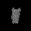 33519MC 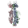 7xy1C 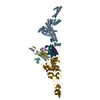 7y1cC 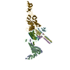 7y22C 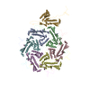 7y23C 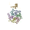 7y3tC 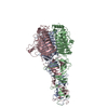 7y5sC M: map data used to model this data C: citing same article ( |
|---|


-Links
 PDBj
PDBj- Assembly
| Deposited unit | 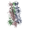
|
|---|---|
| 1 |
|
-Components
| #1: Protein | Mass: 61210.270 Da / Num. of mol.: 3 Source method: isolated from a genetically manipulated source Source: (gene. exp.) Klebsiella phage Kp7 (virus) / Production host:  |
|---|
-Experimental details
-Experiment
| Experiment | Method: ELECTRON MICROSCOPY |
|---|---|
| EM experiment | Aggregation state: PARTICLE / 3D reconstruction method: single particle reconstruction |
- Sample preparation
| Component | Name: Klebsiella phage Kp7 type II tail fiber / Type: COMPLEX / Details: Trimer of tail fiber proteins / Entity ID: all / Source: RECOMBINANT |
|---|---|
| Molecular weight | Experimental value: NO |
| Source (natural) | Organism: Klebsiella phage Kp7 (virus) |
| Source (recombinant) | Organism: |
| Buffer solution | pH: 7.5 |
| Specimen | Embedding applied: NO / Shadowing applied: NO / Staining applied: NO / Vitrification applied: YES |
| Specimen support | Grid material: COPPER / Grid mesh size: 200 divisions/in. / Grid type: Quantifoil R1.2/1.3 |
| Vitrification | Cryogen name: ETHANE |
- Electron microscopy imaging
| Experimental equipment |  Model: Talos Arctica / Image courtesy: FEI Company |
|---|---|
| Microscopy | Model: FEI TALOS ARCTICA |
| Electron gun | Electron source:  FIELD EMISSION GUN / Accelerating voltage: 200 kV / Illumination mode: OTHER FIELD EMISSION GUN / Accelerating voltage: 200 kV / Illumination mode: OTHER |
| Electron lens | Mode: BRIGHT FIELD / Nominal magnification: 36000 X / Nominal defocus max: 1800 nm / Nominal defocus min: 1200 nm / Cs: 2.7 mm / C2 aperture diameter: 50 µm |
| Image recording | Electron dose: 50 e/Å2 / Detector mode: COUNTING / Film or detector model: GATAN K2 SUMMIT (4k x 4k) |
| Image scans | Width: 3710 / Height: 3710 |
- Processing
| EM software | Name: RELION / Version: 3.0.8 / Category: 3D reconstruction |
|---|---|
| CTF correction | Type: PHASE FLIPPING ONLY |
| Symmetry | Point symmetry: C3 (3 fold cyclic) |
| 3D reconstruction | Resolution: 3.8 Å / Resolution method: FSC 0.143 CUT-OFF / Num. of particles: 113834 / Symmetry type: POINT |
| Atomic model building | B value: 146 / Protocol: AB INITIO MODEL / Space: REAL |
