 Movie
Movie Controller

[English] 日本語
 Yorodumi
- EMDB-33519: CryoEM structure of Klebsiella phage Kp7 type II tail fiber gp52 ... -
+ Open data

- Basic information
| Entry |  | |||||||||
|---|---|---|---|---|---|---|---|---|---|---|
| Title | CryoEM structure of Klebsiella phage Kp7 type II tail fiber gp52 in vitro | |||||||||
 Map data Map data | ||||||||||
 Sample Sample |
| |||||||||
 Keywords Keywords | Phage type II tail fiber / VIRAL PROTEIN | |||||||||
| Biological species |  Klebsiella phage Kp7 (virus) Klebsiella phage Kp7 (virus) | |||||||||
| Method | single particle reconstruction / cryo EM / Resolution: 3.8 Å | |||||||||
 Authors Authors | Huang L / Xiang Y | |||||||||
| Funding support |  China, 1 items China, 1 items
| |||||||||
 Citation Citation | Journal: To Be Published Title: Structure and assembly of the Klebsiella pneumoniae phage tail fibers Authors: Huang L / Xiang Y | |||||||||
| History |
|
- Structure visualization
| Supplemental images |
|---|

- Downloads & links
-EMDB archive
| Map data | emd_33519.map.gz | 2.8 MB |  EMDB map data format EMDB map data format | |
|---|---|---|---|---|
| Header (meta data) | emd-33519-v30.xmlemd-33519.xml | 10 KB 10 KB | Display Display | EMDB header |
| FSC (resolution estimation) | emd_33519_fsc.xml | 7.2 KB | Display | FSC data file |
| Images | 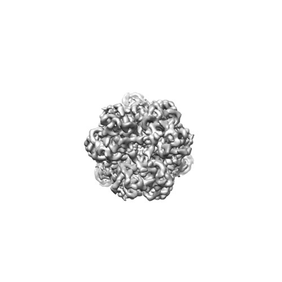 emd_33519.png emd_33519.png | 37.2 KB | ||
| Filedesc metadata | emd-33519.cif.gz | 5.3 KB | ||
| Archive directory |  http://ftp.pdbj.org/pub/emdb/structures/EMD-33519ftp://ftp.pdbj.org/pub/emdb/structures/EMD-33519 http://ftp.pdbj.org/pub/emdb/structures/EMD-33519ftp://ftp.pdbj.org/pub/emdb/structures/EMD-33519 | HTTPS FTP |
-Related structure data
| Related structure data | 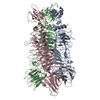 7xycMC 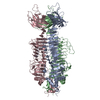 7xy1C 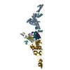 7y1cC 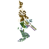 7y22C 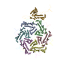 7y23C 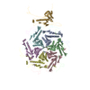 7y3tC 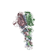 7y5sC M: atomic model generated by this map C: citing same article ( |
|---|


-Links
| EMDB pages | EMDB (EBI/PDBe) / EMDataResource |
|---|
-Map
| File | Download / File: emd_33519.map.gz / Format: CCP4 / Size: 30.5 MB / Type: IMAGE STORED AS FLOATING POINT NUMBER (4 BYTES) | ||||||||||||||||||||||||||||||||||||
|---|---|---|---|---|---|---|---|---|---|---|---|---|---|---|---|---|---|---|---|---|---|---|---|---|---|---|---|---|---|---|---|---|---|---|---|---|---|
| Projections & slices | Image control
Images are generated by Spider. | ||||||||||||||||||||||||||||||||||||
| Voxel size | X=Y=Z: 1.17 Å | ||||||||||||||||||||||||||||||||||||
| Density |
| ||||||||||||||||||||||||||||||||||||
| Symmetry | Space group: 1 | ||||||||||||||||||||||||||||||||||||
| Details | EMDB XML:
|
 Z (Sec.)
 Y (Row.)
 X (Col.)


-Supplemental data
- Sample components
-Entire : Klebsiella phage Kp7 type II tail fiber
| Entire | Name: Klebsiella phage Kp7 type II tail fiber |
|---|---|
| Components |
|
-Supramolecule #1: Klebsiella phage Kp7 type II tail fiber
| Supramolecule | Name: Klebsiella phage Kp7 type II tail fiber / type: complex / ID: 1 / Parent: 0 / Macromolecule list: all / Details: Trimer of tail fiber proteins |
|---|---|
| Source (natural) | Organism: Klebsiella phage Kp7 (virus) |
-Macromolecule #1: phage tail fiber
| Macromolecule | Name: phage tail fiber / type: protein_or_peptide / ID: 1 / Number of copies: 3 / Enantiomer: LEVO |
|---|---|
| Source (natural) | Organism: Klebsiella phage Kp7 (virus) |
| Molecular weight | Theoretical: 61.21027 KDa |
| Recombinant expression | Organism:  |
| Sequence | String: MALVKATFVK DVDGQPWRFS SVAKMKAFNY SCYLGSSVFL ESWHEGAGLG SGLFKVSKGT TEVGDDGSVI VAADGTRLIR VFDGPIFAD MWGALPSSTY NSLPAIKSAY LYASSKLQQL FLGGGSYKVT GSSGIDIDPS LAGISALSRA RVDTTEFTGD Y LFTITSSY ...String: MALVKATFVK DVDGQPWRFS SVAKMKAFNY SCYLGSSVFL ESWHEGAGLG SGLFKVSKGT TEVGDDGSVI VAADGTRLIR VFDGPIFAD MWGALPSSTY NSLPAIKSAY LYASSKLQQL FLGGGSYKVT GSSGIDIDPS LAGISALSRA RVDTTEFTGD Y LFTITSSY SYTPAPYYNN LSVALEGLYV FGNKTEGRSG LLTGRRTTDG VKSYNGQTEI RNCTFDKFDY NIRMGHNSWR IV FYKVNSL NALNANGILY VPSGLDDSGE ILTFYHCQFF DGAGSNIRIS CSSFAMTFVS CSFLNITFTI DAGSSVSVTA LGC NFENPG SQSTRRYIEI TAGHTNIFNV VGGSIVTNAN AGQTQALIHV SANNQINLSN LTIPYGAHYQ QEADSGYHAF CSGQ GYVST SNCSLQLLNG AGCCPIHPSL SVFTNWNLSY ANLNAWTVDK GSAPTSVAEY LSAQGPKGEG VLHVAPTTQG VNISQ VATV SKQAGSMSMS VMVNIISASS NAGQISLAYL DAFDNNLGGV SANLGTTTGW KVIGKNTLRG RLPVGTAKVR LNIQTV AGA DVQYTNILCN II |
-Experimental details
-Structure determination
| Method | cryo EM |
|---|---|
 Processing Processing | single particle reconstruction |
| Aggregation state | particle |
-Sample preparation
| Buffer | pH: 7.5 |
|---|---|
| Grid | Model: Quantifoil R1.2/1.3 / Material: COPPER / Mesh: 200 |
| Vitrification | Cryogen name: ETHANE |
- Electron microscopy
| Microscope | FEI TALOS ARCTICA |
|---|---|
| Image recording | Film or detector model: GATAN K2 SUMMIT (4k x 4k) / Detector mode: COUNTING / Digitization - Dimensions - Width: 3710 pixel / Digitization - Dimensions - Height: 3710 pixel / Average electron dose: 50.0 e/Å2 |
| Electron beam | Acceleration voltage: 200 kV / Electron source:  FIELD EMISSION GUN FIELD EMISSION GUN |
| Electron optics | C2 aperture diameter: 50.0 µm / Illumination mode: OTHER / Imaging mode: BRIGHT FIELD / Cs: 2.7 mm / Nominal defocus max: 1.8 µm / Nominal defocus min: 1.2 µm / Nominal magnification: 36000 |
| Experimental equipment |  Model: Talos Arctica / Image courtesy: FEI Company |
+Image processing

-Atomic model buiding 1
| Refinement | Space: REAL / Protocol: AB INITIO MODEL / Overall B value: 146 |
|---|---|
| Output model | PDB-7xyc: |
