 Movie
Movie Controller

[English] 日本語
 Yorodumi
- EMDB-33623: CryoEM structure of Klebsiella phage Kp7 type I tail fiber gp51 i... -
+ Open data

- Basic information
| Entry |  | |||||||||
|---|---|---|---|---|---|---|---|---|---|---|
| Title | CryoEM structure of Klebsiella phage Kp7 type I tail fiber gp51 in vitro | |||||||||
 Map data Map data | ||||||||||
 Sample Sample |
| |||||||||
 Keywords Keywords | Phage type I tail fiber / VIRAL PROTEIN | |||||||||
| Biological species |  Klebsiella phage Kp7 (virus) Klebsiella phage Kp7 (virus) | |||||||||
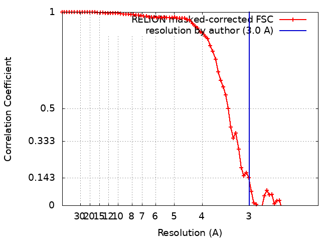
| Method | single particle reconstruction / cryo EM / Resolution: 3.0 Å | |||||||||
 Authors Authors | Huang L / Xiang Y | |||||||||
| Funding support |  China, 1 items China, 1 items
| |||||||||
 Citation Citation | Journal: To Be Published Title: Structure and assembly of the Klebsiella pneumoniae phage tail fibers Authors: Huang L / Xiang Y | |||||||||
| History |
|
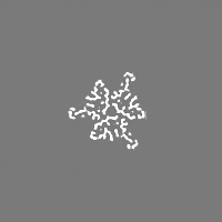
- Structure visualization
| Supplemental images |
|---|

- Downloads & links
-EMDB archive
| Map data | emd_33623.map.gz | 26.9 MB |  EMDB map data format EMDB map data format | |
|---|---|---|---|---|
| Header (meta data) | emd-33623-v30.xmlemd-33623.xml | 13.8 KB 13.8 KB | Display Display | EMDB header |
| FSC (resolution estimation) | emd_33623_fsc.xml | 7.1 KB | Display | FSC data file |
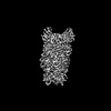
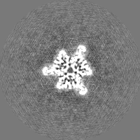
| Images | 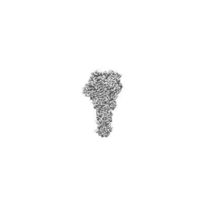 emd_33623.png emd_33623.png | 19.3 KB | ||
| Filedesc metadata | emd-33623.cif.gz | 5.5 KB | ||
| Others | emd_33623_half_map_1.map.gzemd_33623_half_map_2.map.gz | 23.5 MB 23.5 MB | ||
| Archive directory |  http://ftp.pdbj.org/pub/emdb/structures/EMD-33623ftp://ftp.pdbj.org/pub/emdb/structures/EMD-33623 http://ftp.pdbj.org/pub/emdb/structures/EMD-33623ftp://ftp.pdbj.org/pub/emdb/structures/EMD-33623 | HTTPS FTP |
-Related structure data
| Related structure data | 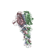 7y5sMC 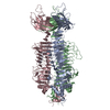 7xy1C 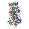 7xycC  7y1cC  7y22C  7y23C  7y3tC M: atomic model generated by this map C: citing same article ( |
|---|


-Links
| EMDB pages | EMDB (EBI/PDBe) / EMDataResource |
|---|
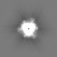
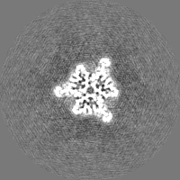
-Map
| File | Download / File: emd_33623.map.gz / Format: CCP4 / Size: 30.5 MB / Type: IMAGE STORED AS FLOATING POINT NUMBER (4 BYTES) | ||||||||||||||||||||||||||||||||||||
|---|---|---|---|---|---|---|---|---|---|---|---|---|---|---|---|---|---|---|---|---|---|---|---|---|---|---|---|---|---|---|---|---|---|---|---|---|---|
| Projections & slices | Image control
Images are generated by Spider. | ||||||||||||||||||||||||||||||||||||
| Voxel size | X=Y=Z: 1.0979 Å | ||||||||||||||||||||||||||||||||||||
| Density |
| ||||||||||||||||||||||||||||||||||||
| Symmetry | Space group: 1 | ||||||||||||||||||||||||||||||||||||
| Details | EMDB XML:
|
 Z (Sec.)
 Y (Row.)
 X (Col.)


-Supplemental data
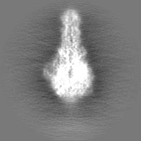
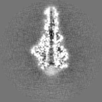
-Half map: #1
| File | emd_33623_half_map_1.map | ||||||||||||
|---|---|---|---|---|---|---|---|---|---|---|---|---|---|
| Projections & Slices |
| ||||||||||||
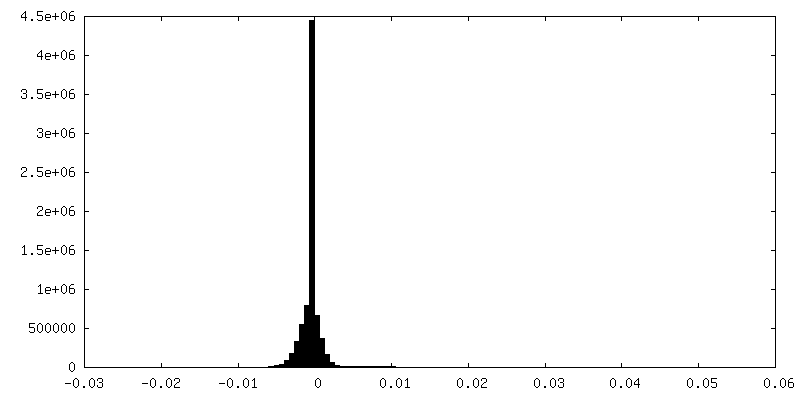
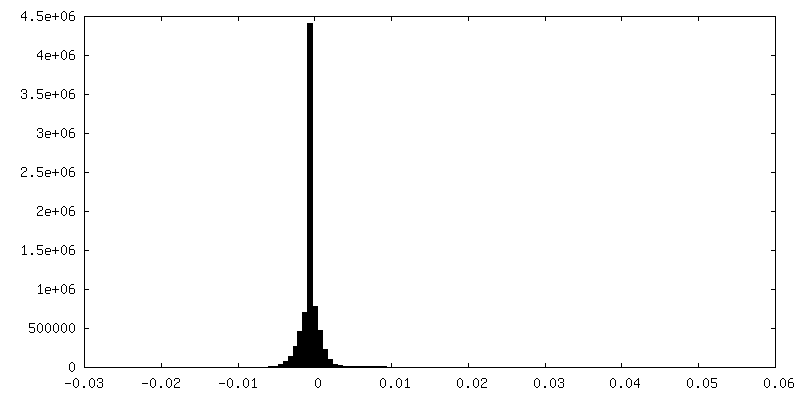
| Density Histograms |

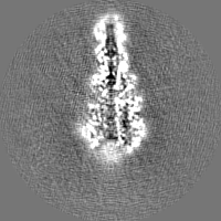

-Half map: #2
| File | emd_33623_half_map_2.map | ||||||||||||
|---|---|---|---|---|---|---|---|---|---|---|---|---|---|
| Projections & Slices |
| ||||||||||||
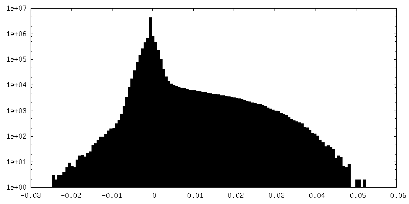
| Density Histograms |


- Sample components
-Entire : Klebsiella phage Kp7 type I tail fiber
| Entire | Name: Klebsiella phage Kp7 type I tail fiber |
|---|---|
| Components |
|
-Supramolecule #1: Klebsiella phage Kp7 type I tail fiber
| Supramolecule | Name: Klebsiella phage Kp7 type I tail fiber / type: complex / ID: 1 / Parent: 0 / Macromolecule list: all / Details: Trimer of tail fiber proteins |
|---|---|
| Source (natural) | Organism: Klebsiella phage Kp7 (virus) |
-Macromolecule #1: phage tail fiber
| Macromolecule | Name: phage tail fiber / type: protein_or_peptide / ID: 1 / Number of copies: 3 / Enantiomer: LEVO |
|---|---|
| Source (natural) | Organism: Klebsiella phage Kp7 (virus) |
| Molecular weight | Theoretical: 73.019125 KDa |
| Recombinant expression | Organism:  |
| Sequence | String: MSVTRLTSSL VRWKSKLANA IQRSLSSKME ESLSVLDFGA VADYNPTTKT GTDNTQAFRN AVAAAIAQNI RNVYAPGGPS AYMTTGEIN LGGEGFTGGE GSRDVWRGIT QGVHFFGDGP YSTIIAFNPP NTDAPCFSAR GGWGTHSPRA LSKLAIEPVN W ADYNATSS ...String: MSVTRLTSSL VRWKSKLANA IQRSLSSKME ESLSVLDFGA VADYNPTTKT GTDNTQAFRN AVAAAIAQNI RNVYAPGGPS AYMTTGEIN LGGEGFTGGE GSRDVWRGIT QGVHFFGDGP YSTIIAFNPP NTDAPCFSAR GGWGTHSPRA LSKLAIEPVN W ADYNATSS GTGVLLQGCC FVPVTDVHIG RFHRGIHFWN KLQGTDDPTN TFTKGDFTEF NRITRVRVFN CDIDVDYQVS LG NNSFHGN SFTDCMCQIN SYGGIGMRMW DDGSRNAIRP SSLPYEYIAN VYNNKHEINW FGSDARTCYL MHIDKAQGRG CNG DMTVEA AVTLRAIGQY WYQSFGSLHS ISAINTVVDG DTDTATRPVA FMWMNSAYPQ VNFDGTDPLL TSGLTPRQYD LNNS GNTGM ELLNIRGANT GAIWSIQNGA ALGWILGRRA QADSRKGTRS VWQFSYNGEV IKSVSAANVG LQNSTGAGFG MLGDT LLRP YAASTISLGS PTYPFTRLRT TDWTVDTNGI VPVQDGIKNI GSSSLRVGTV FAATGTINTS DARLKTEVRP FTSEEI LAA KALSEEIGMY QWVASIESKG DNAREHCGLT VQRAMEVMQQ HGLDPFNYGF ICHDVWDEVV EVSDETGEVI SSTPAGD RY SFRFDQLSLF ISRGIEARMS ELESRM |
-Experimental details
-Structure determination
| Method | cryo EM |
|---|---|
 Processing Processing | single particle reconstruction |
| Aggregation state | particle |
-Sample preparation
| Buffer | pH: 7.5 |
|---|---|
| Grid | Model: Quantifoil R1.2/1.3 / Material: COPPER / Mesh: 200 |
| Vitrification | Cryogen name: ETHANE |
- Electron microscopy
| Microscope | FEI TITAN KRIOS |
|---|---|
| Image recording | Film or detector model: GATAN K3 (6k x 4k) / Digitization - Dimensions - Width: 5760 pixel / Digitization - Dimensions - Height: 4092 pixel / Average electron dose: 50.0 e/Å2 |
| Electron beam | Acceleration voltage: 300 kV / Electron source:  FIELD EMISSION GUN FIELD EMISSION GUN |
| Electron optics | C2 aperture diameter: 50.0 µm / Illumination mode: FLOOD BEAM / Imaging mode: BRIGHT FIELD / Cs: 2.7 mm / Nominal defocus max: 1.5 µm / Nominal defocus min: 1.2 µm |
| Experimental equipment |  Model: Titan Krios / Image courtesy: FEI Company |
+Image processing
-Atomic model buiding 1
| Refinement | Protocol: AB INITIO MODEL |
|---|---|
| Output model | PDB-7y5s: |
