 ムービー
ムービー コントローラー


+ データを開く

- 基本情報
基本情報
| 登録情報 | データベース: PDB / ID: 7xv2 | ||||||
|---|---|---|---|---|---|---|---|
| タイトル | TRIM E3 ubiquitin ligase | ||||||
 要素 要素 | Tripartite motif-containing protein 72 | ||||||
 キーワード キーワード | MEMBRANE PROTEIN / TRIM / Tripartite motif / Ubiquitin ligase / Coiled coil / B-box / PRY-SPRY / LIGASE / METAL BINDING PROTEIN | ||||||
| 機能・相同性 |  機能・相同性情報 機能・相同性情報muscle system process / negative regulation of insulin-like growth factor receptor signaling pathway / negative regulation of myotube differentiation / plasma membrane repair / vesicle budding from membrane / ubiquitin conjugating enzyme binding / mitogen-activated protein kinase kinase kinase binding / muscle organ development / phosphatidylserine binding / exocytosis ...muscle system process / negative regulation of insulin-like growth factor receptor signaling pathway / negative regulation of myotube differentiation / plasma membrane repair / vesicle budding from membrane / ubiquitin conjugating enzyme binding / mitogen-activated protein kinase kinase kinase binding / muscle organ development / phosphatidylserine binding / exocytosis / negative regulation of insulin receptor signaling pathway / cytoplasmic vesicle membrane / protein homooligomerization / sarcolemma / RING-type E3 ubiquitin transferase / ubiquitin protein ligase activity / proteasome-mediated ubiquitin-dependent protein catabolic process / zinc ion binding / identical protein binding 類似検索 - 分子機能 | ||||||
| 生物種 |  | ||||||
| 手法 |  X線回折 / シンクロトロン / 分子置換 / 解像度: 2.75 Å X線回折 / シンクロトロン / 分子置換 / 解像度: 2.75 Å | ||||||
 データ登録者 データ登録者 | Park, S.H. / Song, H.K. | ||||||
| 資金援助 |  韓国, 1件 韓国, 1件
| ||||||
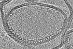
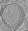
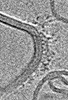
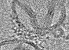
 引用 引用 | ジャーナル: Nat Struct Mol Biol / 年: 2023 タイトル: Structure and activation of the RING E3 ubiquitin ligase TRIM72 on the membrane. 著者: Si Hoon Park / Juhyun Han / Byung-Cheon Jeong / Ju Han Song / Se Hwan Jang / Hyeongseop Jeong / Bong Heon Kim / Young-Gyu Ko / Zee-Yong Park / Kyung Eun Lee / Jaekyung Hyun / Hyun Kyu Song /   要旨: Defects in plasma membrane repair can lead to muscle and heart diseases in humans. Tripartite motif-containing protein (TRIM)72 (mitsugumin 53; MG53) has been determined to rapidly nucleate vesicles ...Defects in plasma membrane repair can lead to muscle and heart diseases in humans. Tripartite motif-containing protein (TRIM)72 (mitsugumin 53; MG53) has been determined to rapidly nucleate vesicles at the site of membrane damage, but the underlying molecular mechanisms remain poorly understood. Here we present the structure of Mus musculus TRIM72, a complete model of a TRIM E3 ubiquitin ligase. We demonstrated that the interaction between TRIM72 and phosphatidylserine-enriched membranes is necessary for its oligomeric assembly and ubiquitination activity. Using cryogenic electron tomography and subtomogram averaging, we elucidated a higher-order model of TRIM72 assembly on the phospholipid bilayer. Combining structural and biochemical techniques, we developed a working molecular model of TRIM72, providing insights into the regulation of RING-type E3 ligases through the cooperation of multiple domains in higher-order assemblies. Our findings establish a fundamental basis for the study of TRIM E3 ligases and have therapeutic implications for diseases associated with membrane repair. | ||||||
| 履歴 |
|
- 構造の表示
構造の表示
| 構造ビューア | 分子: MolmilJmol/JSmol |
|---|
- ダウンロードとリンク
-ダウンロード
| PDBx/mmCIF形式 | 7xv2.cif.gz | 201.9 KB | 表示 | PDBx/mmCIF形式 |
|---|---|---|---|---|
| PDB形式 | pdb7xv2.ent.gz | 134.9 KB | 表示 | PDB形式 |
| PDBx/mmJSON形式 | 7xv2.json.gz | ツリー表示 | PDBx/mmJSON形式 | |
| その他 |  その他のダウンロード その他のダウンロード |
-検証レポート
| 文書・要旨 | 7xv2_validation.pdf.gz | 911.9 KB | 表示 | wwPDB検証レポート |
|---|---|---|---|---|
| 文書・詳細版 | 7xv2_full_validation.pdf.gz | 918.6 KB | 表示 | |
| XML形式データ | 7xv2_validation.xml.gz | 15.5 KB | 表示 | |
| CIF形式データ | 7xv2_validation.cif.gz | 20.3 KB | 表示 | |
| アーカイブディレクトリ | https://data.pdbj.org/pub/pdb/validation_reports/xv/7xv2ftp://data.pdbj.org/pub/pdb/validation_reports/xv/7xv2 | HTTPS FTP |
-関連構造データ
| 関連構造データ |  7xyyC  7xyzC 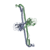 7xz0C 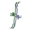 7xz1C 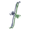 7xz2C 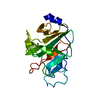 3kb5S S: 精密化の開始モデル C: 同じ文献を引用 ( |
|---|---|
| 類似構造データ | |
| その他のデータベース |
|


-リンク
 PDBj
PDBj

- 集合体
集合体
| 登録構造単位 | 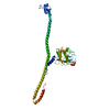
| ||||||||||||
|---|---|---|---|---|---|---|---|---|---|---|---|---|---|
| 1 | 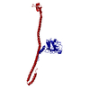
| ||||||||||||
| 単位格子 |
|
-要素
| #1: タンパク質 | 分子量: 44200.523 Da / 分子数: 1 / 変異: C144S,C242S,K279H,A283H / 由来タイプ: 組換発現 / 由来: (組換発現)  | ||
|---|---|---|---|
| #2: 化合物 |   分子量: 65.409 Da / 分子数: 2 / 由来タイプ: 合成 / 式: Zn / タイプ: SUBJECT OF INVESTIGATION 分子量: 65.409 Da / 分子数: 2 / 由来タイプ: 合成 / 式: Zn / タイプ: SUBJECT OF INVESTIGATION研究の焦点であるリガンドがあるか | Y | |
-実験情報
-実験
| 実験 | 手法: X線回折 / 使用した結晶の数: 1 |
|---|
- 試料調製
試料調製
| 結晶 | マシュー密度: 4.3 Å3/Da / 溶媒含有率: 71.42 % |
|---|---|
| 結晶化 | 温度: 277 K / 手法: 蒸気拡散法, ハンギングドロップ法 / pH: 8.5 / 詳細: 100 mM Tris-HCl pH 8.5, 30% (v/v) PEG 400 |
-データ収集
| 回折 | 平均測定温度: 100 K / Serial crystal experiment: N |
|---|---|
| 放射光源 | 由来: シンクロトロン / サイト: PAL/PLS / ビームライン: 5C (4A) / 波長: 0.9796 Å |
| 検出器 | タイプ: RAYONIX MX-225 / 検出器: CCD / 日付: 2014年10月11日 |
| 放射 | プロトコル: SINGLE WAVELENGTH / 単色(M)・ラウエ(L): M / 散乱光タイプ: x-ray |
| 放射波長 | 波長: 0.9796 Å / 相対比: 1 |
| 反射 | 解像度: 2.75→30 Å / Num. obs: 19188 / % possible obs: 97.6 % / 冗長度: 4.1 % / Biso Wilson estimate: 80.36 Å2 / CC1/2: 0.99 / CC star: 0.997 / Rmerge(I) obs: 0.087 / Net I/σ(I): 27.3 |
| 反射 シェル | 解像度: 2.75→2.8 Å / Mean I/σ(I) obs: 1.3 / Num. unique obs: 948 / CC1/2: 0.806 / CC star: 0.945 / % possible all: 98.9 |
- 解析
解析
| ソフトウェア |
| |||||||||||||||||||||||||||||||||||||||||||||||||||||||||||||||||||||||||||||||||||||||||||||||||||||||||||||||||||||||||||||
|---|---|---|---|---|---|---|---|---|---|---|---|---|---|---|---|---|---|---|---|---|---|---|---|---|---|---|---|---|---|---|---|---|---|---|---|---|---|---|---|---|---|---|---|---|---|---|---|---|---|---|---|---|---|---|---|---|---|---|---|---|---|---|---|---|---|---|---|---|---|---|---|---|---|---|---|---|---|---|---|---|---|---|---|---|---|---|---|---|---|---|---|---|---|---|---|---|---|---|---|---|---|---|---|---|---|---|---|---|---|---|---|---|---|---|---|---|---|---|---|---|---|---|---|---|---|---|
| 精密化 | 構造決定の手法: 分子置換 開始モデル: 3KB5 解像度: 2.75→27.41 Å / SU ML: 0.4255 / 交差検証法: FREE R-VALUE / σ(F): 1.35 / 位相誤差: 32.2552 立体化学のターゲット値: GeoStd + Monomer Library + CDL v1.2
| |||||||||||||||||||||||||||||||||||||||||||||||||||||||||||||||||||||||||||||||||||||||||||||||||||||||||||||||||||||||||||||
| 溶媒の処理 | 減衰半径: 0.9 Å / VDWプローブ半径: 1.11 Å / 溶媒モデル: FLAT BULK SOLVENT MODEL | |||||||||||||||||||||||||||||||||||||||||||||||||||||||||||||||||||||||||||||||||||||||||||||||||||||||||||||||||||||||||||||
| 原子変位パラメータ | Biso mean: 115.04 Å2 | |||||||||||||||||||||||||||||||||||||||||||||||||||||||||||||||||||||||||||||||||||||||||||||||||||||||||||||||||||||||||||||
| 精密化ステップ | サイクル: LAST / 解像度: 2.75→27.41 Å
| |||||||||||||||||||||||||||||||||||||||||||||||||||||||||||||||||||||||||||||||||||||||||||||||||||||||||||||||||||||||||||||
| 拘束条件 |
| |||||||||||||||||||||||||||||||||||||||||||||||||||||||||||||||||||||||||||||||||||||||||||||||||||||||||||||||||||||||||||||
| LS精密化 シェル |
| |||||||||||||||||||||||||||||||||||||||||||||||||||||||||||||||||||||||||||||||||||||||||||||||||||||||||||||||||||||||||||||
| 精密化 TLS | 手法: refined / Refine-ID: X-RAY DIFFRACTION
| |||||||||||||||||||||||||||||||||||||||||||||||||||||||||||||||||||||||||||||||||||||||||||||||||||||||||||||||||||||||||||||
| 精密化 TLSグループ |
|
