 Movie
Movie Controller


+ Open data

- Basic information
| Entry | Database: PDB / ID: 7xv2 | ||||||
|---|---|---|---|---|---|---|---|
| Title | TRIM E3 ubiquitin ligase | ||||||
 Components Components | Tripartite motif-containing protein 72 | ||||||
 Keywords Keywords | MEMBRANE PROTEIN / TRIM / Tripartite motif / Ubiquitin ligase / Coiled coil / B-box / PRY-SPRY / LIGASE / METAL BINDING PROTEIN | ||||||
| Function / homology |  Function and homology information Function and homology informationmuscle system process / negative regulation of insulin-like growth factor receptor signaling pathway / negative regulation of myotube differentiation / vesicle budding from membrane / plasma membrane repair / mitogen-activated protein kinase kinase kinase binding / ubiquitin conjugating enzyme binding / muscle organ development / phosphatidylserine binding / exocytosis ...muscle system process / negative regulation of insulin-like growth factor receptor signaling pathway / negative regulation of myotube differentiation / vesicle budding from membrane / plasma membrane repair / mitogen-activated protein kinase kinase kinase binding / ubiquitin conjugating enzyme binding / muscle organ development / phosphatidylserine binding / exocytosis / negative regulation of insulin receptor signaling pathway / cytoplasmic vesicle membrane / protein homooligomerization / sarcolemma / RING-type E3 ubiquitin transferase / ubiquitin protein ligase activity / proteasome-mediated ubiquitin-dependent protein catabolic process / innate immune response / zinc ion binding / identical protein binding / cytoplasm Similarity search - Function | ||||||
| Biological species |  | ||||||
| Method |  X-RAY DIFFRACTION / SYNCHROTRON / MOLECULAR REPLACEMENT / Resolution: 2.75 Å X-RAY DIFFRACTION / SYNCHROTRON / MOLECULAR REPLACEMENT / Resolution: 2.75 Å | ||||||
 Authors Authors | Park, S.H. / Song, H.K. | ||||||
| Funding support |  Korea, Republic Of, 1items Korea, Republic Of, 1items
| ||||||
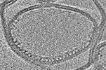
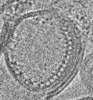
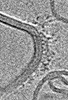
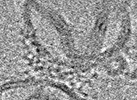
 Citation Citation | Journal: Nat Struct Mol Biol / Year: 2023 Title: Structure and activation of the RING E3 ubiquitin ligase TRIM72 on the membrane. Authors: Si Hoon Park / Juhyun Han / Byung-Cheon Jeong / Ju Han Song / Se Hwan Jang / Hyeongseop Jeong / Bong Heon Kim / Young-Gyu Ko / Zee-Yong Park / Kyung Eun Lee / Jaekyung Hyun / Hyun Kyu Song /   Abstract: Defects in plasma membrane repair can lead to muscle and heart diseases in humans. Tripartite motif-containing protein (TRIM)72 (mitsugumin 53; MG53) has been determined to rapidly nucleate vesicles ...Defects in plasma membrane repair can lead to muscle and heart diseases in humans. Tripartite motif-containing protein (TRIM)72 (mitsugumin 53; MG53) has been determined to rapidly nucleate vesicles at the site of membrane damage, but the underlying molecular mechanisms remain poorly understood. Here we present the structure of Mus musculus TRIM72, a complete model of a TRIM E3 ubiquitin ligase. We demonstrated that the interaction between TRIM72 and phosphatidylserine-enriched membranes is necessary for its oligomeric assembly and ubiquitination activity. Using cryogenic electron tomography and subtomogram averaging, we elucidated a higher-order model of TRIM72 assembly on the phospholipid bilayer. Combining structural and biochemical techniques, we developed a working molecular model of TRIM72, providing insights into the regulation of RING-type E3 ligases through the cooperation of multiple domains in higher-order assemblies. Our findings establish a fundamental basis for the study of TRIM E3 ligases and have therapeutic implications for diseases associated with membrane repair. | ||||||
| History |
|
- Structure visualization
| Structure viewer | Molecule: MolmilJmol/JSmol |
|---|
- Downloads & links
-Download
| PDBx/mmCIF format | 7xv2.cif.gz | 201.9 KB | Display | PDBx/mmCIF format |
|---|---|---|---|---|
| PDB format | pdb7xv2.ent.gz | 134.9 KB | Display | PDB format |
| PDBx/mmJSON format | 7xv2.json.gz | Tree view | PDBx/mmJSON format | |
| Others |  Other downloads Other downloads |
-Validation report
| Arichive directory | https://data.pdbj.org/pub/pdb/validation_reports/xv/7xv2ftp://data.pdbj.org/pub/pdb/validation_reports/xv/7xv2 | HTTPS FTP |
|---|
-Related structure data
| Related structure data |  7xyyC  7xyzC 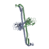 7xz0C 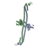 7xz1C 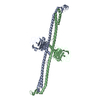 7xz2C 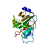 3kb5S S: Starting model for refinement C: citing same article ( |
|---|---|
| Similar structure data | |
| Other databases |
|


-Links
 PDBj
PDBj

- Assembly
| Deposited unit | 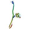
| ||||||||||||
|---|---|---|---|---|---|---|---|---|---|---|---|---|---|
| 1 | 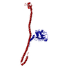
| ||||||||||||
| Unit cell |
|
-Components
| #1: Protein | Mass: 44200.523 Da / Num. of mol.: 1 / Mutation: C144S,C242S,K279H,A283H Source method: isolated from a genetically manipulated source Source: (gene. exp.)  | ||
|---|---|---|---|
| #2: Chemical |   Mass: 65.409 Da / Num. of mol.: 2 / Source method: obtained synthetically / Formula: Zn / Feature type: SUBJECT OF INVESTIGATION Mass: 65.409 Da / Num. of mol.: 2 / Source method: obtained synthetically / Formula: Zn / Feature type: SUBJECT OF INVESTIGATIONHas ligand of interest | Y | |
-Experimental details
-Experiment
| Experiment | Method: X-RAY DIFFRACTION / Number of used crystals: 1 |
|---|
- Sample preparation
| Crystal | Density Matthews: 4.3 Å3/Da / Density % sol: 71.42 % |
|---|---|
| Crystal grow | Temperature: 277 K / Method: vapor diffusion, hanging drop / pH: 8.5 / Details: 100 mM Tris-HCl pH 8.5, 30% (v/v) PEG 400 |
-Data collection
| Diffraction | Mean temperature: 100 K / Serial crystal experiment: N |
|---|---|
| Diffraction source | Source: SYNCHROTRON / Site: PAL/PLS / Beamline: 5C (4A) / Wavelength: 0.9796 Å |
| Detector | Type: RAYONIX MX-225 / Detector: CCD / Date: Oct 11, 2014 |
| Radiation | Protocol: SINGLE WAVELENGTH / Monochromatic (M) / Laue (L): M / Scattering type: x-ray |
| Radiation wavelength | Wavelength: 0.9796 Å / Relative weight: 1 |
| Reflection | Resolution: 2.75→30 Å / Num. obs: 19188 / % possible obs: 97.6 % / Redundancy: 4.1 % / Biso Wilson estimate: 80.36 Å2 / CC1/2: 0.99 / CC star: 0.997 / Rmerge(I) obs: 0.087 / Net I/σ(I): 27.3 |
| Reflection shell | Resolution: 2.75→2.8 Å / Mean I/σ(I) obs: 1.3 / Num. unique obs: 948 / CC1/2: 0.806 / CC star: 0.945 / % possible all: 98.9 |
- Processing
| Software |
| |||||||||||||||||||||||||||||||||||||||||||||||||||||||||||||||||||||||||||||||||||||||||||||||||||||||||||||||||||||||||||||
|---|---|---|---|---|---|---|---|---|---|---|---|---|---|---|---|---|---|---|---|---|---|---|---|---|---|---|---|---|---|---|---|---|---|---|---|---|---|---|---|---|---|---|---|---|---|---|---|---|---|---|---|---|---|---|---|---|---|---|---|---|---|---|---|---|---|---|---|---|---|---|---|---|---|---|---|---|---|---|---|---|---|---|---|---|---|---|---|---|---|---|---|---|---|---|---|---|---|---|---|---|---|---|---|---|---|---|---|---|---|---|---|---|---|---|---|---|---|---|---|---|---|---|---|---|---|---|
| Refinement | Method to determine structure: MOLECULAR REPLACEMENT Starting model: 3KB5 Resolution: 2.75→27.41 Å / SU ML: 0.4255 / Cross valid method: FREE R-VALUE / σ(F): 1.35 / Phase error: 32.2552 Stereochemistry target values: GeoStd + Monomer Library + CDL v1.2
| |||||||||||||||||||||||||||||||||||||||||||||||||||||||||||||||||||||||||||||||||||||||||||||||||||||||||||||||||||||||||||||
| Solvent computation | Shrinkage radii: 0.9 Å / VDW probe radii: 1.11 Å / Solvent model: FLAT BULK SOLVENT MODEL | |||||||||||||||||||||||||||||||||||||||||||||||||||||||||||||||||||||||||||||||||||||||||||||||||||||||||||||||||||||||||||||
| Displacement parameters | Biso mean: 115.04 Å2 | |||||||||||||||||||||||||||||||||||||||||||||||||||||||||||||||||||||||||||||||||||||||||||||||||||||||||||||||||||||||||||||
| Refinement step | Cycle: LAST / Resolution: 2.75→27.41 Å
| |||||||||||||||||||||||||||||||||||||||||||||||||||||||||||||||||||||||||||||||||||||||||||||||||||||||||||||||||||||||||||||
| Refine LS restraints |
| |||||||||||||||||||||||||||||||||||||||||||||||||||||||||||||||||||||||||||||||||||||||||||||||||||||||||||||||||||||||||||||
| LS refinement shell |
| |||||||||||||||||||||||||||||||||||||||||||||||||||||||||||||||||||||||||||||||||||||||||||||||||||||||||||||||||||||||||||||
| Refinement TLS params. | Method: refined / Refine-ID: X-RAY DIFFRACTION
| |||||||||||||||||||||||||||||||||||||||||||||||||||||||||||||||||||||||||||||||||||||||||||||||||||||||||||||||||||||||||||||
| Refinement TLS group |
|
