 Movie
Movie Controller

[English] 日本語
 Yorodumi
- PDB-7x9f: Crystal structure of actinomycin D-echinomycin-d(AGCGCGT/ACGCGCT)... -
+ Open data

- Basic information
| Entry | Database: PDB / ID: 7x9f | ||||||||||||
|---|---|---|---|---|---|---|---|---|---|---|---|---|---|
| Title | Crystal structure of actinomycin D-echinomycin-d(AGCGCGT/ACGCGCT) complex | ||||||||||||
 Components Components |
| ||||||||||||
 Keywords Keywords | DNA/ANTIBIOTIC / G:C canonical DNA / Single-strand DNA twist / four-way junction / Echinomycin / Actinomycin D / DNA / DNA-ANTIBIOTIC complex | ||||||||||||
| Function / homology | Actinomycin D / Echinomycin / : / 2-CARBOXYQUINOXALINE / DNA Function and homology information Function and homology information | ||||||||||||
| Biological species |  Streptomyces (bacteria) Streptomyces (bacteria)synthetic construct (others) | ||||||||||||
| Method |  X-RAY DIFFRACTION / SYNCHROTRON / MOLECULAR REPLACEMENT / Resolution: 2.96 Å X-RAY DIFFRACTION / SYNCHROTRON / MOLECULAR REPLACEMENT / Resolution: 2.96 Å | ||||||||||||
 Authors Authors | Kao, S.H. / Satange, R.B. / Hou, M.H. | ||||||||||||
| Funding support |  Taiwan, 2items Taiwan, 2items
| ||||||||||||
 Citation Citation | Journal: Nucleic Acids Res. / Year: 2022 Title: Staggered intercalation of DNA duplexes with base-pair modulation by two distinct drug molecules induces asymmetric backbone twisting and structure polymorphism. Authors: Satange, R. / Kao, S.H. / Chien, C.M. / Chou, S.H. / Lin, C.C. / Neidle, S. / Hou, M.H. | ||||||||||||
| History |
|
- Structure visualization
| Structure viewer | Molecule: MolmilJmol/JSmol |
|---|
- Downloads & links
-Download
| PDBx/mmCIF format | 7x9f.cif.gz | 61.5 KB | Display | PDBx/mmCIF format |
|---|---|---|---|---|
| PDB format | pdb7x9f.ent.gz | 45.5 KB | Display | PDB format |
| PDBx/mmJSON format | 7x9f.json.gz | Tree view | PDBx/mmJSON format | |
| Others |  Other downloads Other downloads |
-Validation report
| Arichive directory | https://data.pdbj.org/pub/pdb/validation_reports/x9/7x9fftp://data.pdbj.org/pub/pdb/validation_reports/x9/7x9f | HTTPS FTP |
|---|
-Related structure data
| Related structure data | 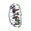 7x6rC  7x97C 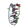 7xdjC 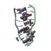 7dq0S S: Starting model for refinement C: citing same article ( |
|---|---|
| Similar structure data |
-Links
 PDBj
PDBj


- Assembly
| Deposited unit | 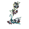
| |||||||||
|---|---|---|---|---|---|---|---|---|---|---|
| 1 | 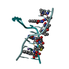
| |||||||||
| 2 | 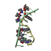
| |||||||||
| Unit cell |
| |||||||||
| Components on special symmetry positions |
|
-Components
-DNA chain , 2 types, 4 molecules FDOB
| #1: DNA chain | Mass: 2098.399 Da / Num. of mol.: 2 / Source method: obtained synthetically / Source: (synth.) synthetic construct (others) #2: DNA chain | Mass: 2138.423 Da / Num. of mol.: 2 / Source method: obtained synthetically / Source: (synth.) synthetic construct (others) |
|---|
-Protein/peptide , 2 types, 4 molecules GCHA
| #3: Protein/peptide | 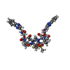  Type: Cyclic depsipeptide / Class: Antibiotic / Mass: 809.008 Da / Num. of mol.: 2 / Source method: isolated from a natural source Type: Cyclic depsipeptide / Class: Antibiotic / Mass: 809.008 Da / Num. of mol.: 2 / Source method: isolated from a natural sourceDetails: ECHINOMYCIN IS A BICYCLIC OCTADEPSIPEPTIDE. BICYCLIZATION IS ACHIEVED BY LINKING THE N- AND THE C- TERMINI, AND A THIOACETAL BOND BETWEEN RESIDUES 3 AND 7. THE TWO QUINOXALINE CHROMOPHORES ...Details: ECHINOMYCIN IS A BICYCLIC OCTADEPSIPEPTIDE. BICYCLIZATION IS ACHIEVED BY LINKING THE N- AND THE C- TERMINI, AND A THIOACETAL BOND BETWEEN RESIDUES 3 AND 7. THE TWO QUINOXALINE CHROMOPHORES ARE LINKED TO THE D-SERINE RESIDUES, RESIDUES 1 AND 5. Source: (natural) Streptomyces (bacteria) / References: Echinomycin#4: Protein/peptide | 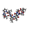  Type: Polypeptide / Class: Antibiotic / Mass: 1291.446 Da / Num. of mol.: 2 / Source method: isolated from a natural source Type: Polypeptide / Class: Antibiotic / Mass: 1291.446 Da / Num. of mol.: 2 / Source method: isolated from a natural sourceDetails: ACTINOMYCIN D CONSISTS OF TWO PENTAMER RINGS LINKED BY THE CHROMOPHORE (PXZ) Source: (natural) Streptomyces (bacteria) / References: Actinomycin D |
|---|
-Non-polymers , 3 types, 12 molecules 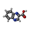


| #5: Chemical | ChemComp-QUI /  Mass: 174.156 Da / Num. of mol.: 4 / Source method: obtained synthetically / Formula: C9H6N2O2 Mass: 174.156 Da / Num. of mol.: 4 / Source method: obtained synthetically / Formula: C9H6N2O2#6: Chemical | ChemComp-MN / |  Mass: 54.938 Da / Num. of mol.: 1 / Source method: obtained synthetically / Formula: Mn Mass: 54.938 Da / Num. of mol.: 1 / Source method: obtained synthetically / Formula: Mn#7: Water | ChemComp-HOH / | Mass: 18.015 Da / Num. of mol.: 7 / Source method: isolated from a natural source / Formula: H2O |
|---|
-Details
| Compound details | THE ECHINOMYCIN IS A BICYCLIC OCTADEPSIPEPTIDE, A MEMBER OF THE QUINOXALINE CLASS OF ANTIBIOTICS. ...THE ECHINOMYCI |
|---|---|
| Has ligand of interest | N |
-Experimental details
-Experiment
| Experiment | Method: X-RAY DIFFRACTION / Number of used crystals: 1 |
|---|
- Sample preparation
| Crystal | Density Matthews: 3.55 Å3/Da / Density % sol: 65.31 % |
|---|---|
| Crystal grow | Temperature: 293 K / Method: vapor diffusion, sitting drop / pH: 6 Details: 10 mM MES buffer (pH 6.0), 5 mM Spermine tetrahydrochloride, 2% PEG200, 3 mM Manganese chloride, 0.25 mM Oligonucleotides, 0.25 mM echinomycin, 0.125 mM actinomycin D |
-Data collection
| Diffraction | Mean temperature: 100 K / Serial crystal experiment: N |
|---|---|
| Diffraction source | Source: SYNCHROTRON / Site: NSRRC / Beamline: BL15A1 / Wavelength: 1 Å |
| Detector | Type: RAYONIX MX-300 / Detector: CCD / Date: Apr 19, 2019 |
| Radiation | Protocol: SINGLE WAVELENGTH / Monochromatic (M) / Laue (L): M / Scattering type: x-ray |
| Radiation wavelength | Wavelength: 1 Å / Relative weight: 1 |
| Reflection | Resolution: 2.96→30 Å / Num. obs: 4205 / % possible obs: 99.9 % / Redundancy: 7.6 % / Biso Wilson estimate: 53.86 Å2 / Rmerge(I) obs: 0.087 / Rpim(I) all: 0.034 / Rrim(I) all: 0.094 / Χ2: 1.196 / Net I/σ(I): 9.2 / Num. measured all: 31951 |
| Reflection shell | Resolution: 2.96→3.07 Å / Redundancy: 8 % / Rmerge(I) obs: 0.75 / Num. unique obs: 475 / CC1/2: 0.999 / Rpim(I) all: 0.012 / Rrim(I) all: 0.032 / Χ2: 1.035 / % possible all: 100 |
- Processing
| Software |
| |||||||||||||||||||||||||||||||||||||||||||||||||||||||||||||||||||||||||||||||||||||||||||||||||||||||||||||||||||||||||||||||||||||||||||||||||||||||||||||||||||||||||||||||||||||||||||||||||||||||||||||||||||||||||||||||||
|---|---|---|---|---|---|---|---|---|---|---|---|---|---|---|---|---|---|---|---|---|---|---|---|---|---|---|---|---|---|---|---|---|---|---|---|---|---|---|---|---|---|---|---|---|---|---|---|---|---|---|---|---|---|---|---|---|---|---|---|---|---|---|---|---|---|---|---|---|---|---|---|---|---|---|---|---|---|---|---|---|---|---|---|---|---|---|---|---|---|---|---|---|---|---|---|---|---|---|---|---|---|---|---|---|---|---|---|---|---|---|---|---|---|---|---|---|---|---|---|---|---|---|---|---|---|---|---|---|---|---|---|---|---|---|---|---|---|---|---|---|---|---|---|---|---|---|---|---|---|---|---|---|---|---|---|---|---|---|---|---|---|---|---|---|---|---|---|---|---|---|---|---|---|---|---|---|---|---|---|---|---|---|---|---|---|---|---|---|---|---|---|---|---|---|---|---|---|---|---|---|---|---|---|---|---|---|---|---|---|---|---|---|---|---|---|---|---|---|---|---|---|---|---|---|---|---|
| Refinement | Method to determine structure: MOLECULAR REPLACEMENT Starting model: 7DQ0 Resolution: 2.96→27.8 Å / SU ML: 0.37 / Cross valid method: THROUGHOUT / σ(F): 1.36 / Phase error: 29.43 / Stereochemistry target values: ML
| |||||||||||||||||||||||||||||||||||||||||||||||||||||||||||||||||||||||||||||||||||||||||||||||||||||||||||||||||||||||||||||||||||||||||||||||||||||||||||||||||||||||||||||||||||||||||||||||||||||||||||||||||||||||||||||||||
| Solvent computation | Shrinkage radii: 0.9 Å / VDW probe radii: 1.11 Å / Solvent model: FLAT BULK SOLVENT MODEL | |||||||||||||||||||||||||||||||||||||||||||||||||||||||||||||||||||||||||||||||||||||||||||||||||||||||||||||||||||||||||||||||||||||||||||||||||||||||||||||||||||||||||||||||||||||||||||||||||||||||||||||||||||||||||||||||||
| Displacement parameters | Biso max: 201.3 Å2 / Biso mean: 76.81 Å2 / Biso min: 35.23 Å2 | |||||||||||||||||||||||||||||||||||||||||||||||||||||||||||||||||||||||||||||||||||||||||||||||||||||||||||||||||||||||||||||||||||||||||||||||||||||||||||||||||||||||||||||||||||||||||||||||||||||||||||||||||||||||||||||||||
| Refinement step | Cycle: final / Resolution: 2.96→27.8 Å
| |||||||||||||||||||||||||||||||||||||||||||||||||||||||||||||||||||||||||||||||||||||||||||||||||||||||||||||||||||||||||||||||||||||||||||||||||||||||||||||||||||||||||||||||||||||||||||||||||||||||||||||||||||||||||||||||||
| LS refinement shell | Refine-ID: X-RAY DIFFRACTION / Rfactor Rfree error: 0 / Total num. of bins used: 3
| |||||||||||||||||||||||||||||||||||||||||||||||||||||||||||||||||||||||||||||||||||||||||||||||||||||||||||||||||||||||||||||||||||||||||||||||||||||||||||||||||||||||||||||||||||||||||||||||||||||||||||||||||||||||||||||||||
| Refinement TLS params. | Method: refined / Refine-ID: X-RAY DIFFRACTION
| |||||||||||||||||||||||||||||||||||||||||||||||||||||||||||||||||||||||||||||||||||||||||||||||||||||||||||||||||||||||||||||||||||||||||||||||||||||||||||||||||||||||||||||||||||||||||||||||||||||||||||||||||||||||||||||||||
| Refinement TLS group |
|
