 Movie
Movie Controller


+ Open data

- Basic information
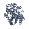
| Entry | Database: PDB / ID: 7wan | ||||||
|---|---|---|---|---|---|---|---|
| Title | Crystal structure of HaloTag complexed with UL2 | ||||||
 Components Components | Haloalkane dehalogenase | ||||||
 Keywords Keywords | HYDROLASE / halotag / haloalkane dehalogenase | ||||||
| Function / homology |  Function and homology information Function and homology informationhaloalkane dehalogenase / haloalkane dehalogenase activity / response to toxic substance / membrane Similarity search - Function | ||||||
| Biological species |  Rhodococcus sp. (bacteria) Rhodococcus sp. (bacteria) | ||||||
| Method |  X-RAY DIFFRACTION / SYNCHROTRON / MOLECULAR REPLACEMENT / Resolution: 2.284 Å X-RAY DIFFRACTION / SYNCHROTRON / MOLECULAR REPLACEMENT / Resolution: 2.284 Å | ||||||
 Authors Authors | Pratyush, M. / Kang, M. / Lee, H. / Lee, C. / Rhee, H. | ||||||
| Funding support | 1items
| ||||||
 Citation Citation | Journal: Chem Sci / Year: 2022 Title: A chemical tool for blue light-inducible proximity photo-crosslinking in live cells. Authors: Mishra, P.K. / Kang, M.G. / Lee, H. / Kim, S. / Choi, S. / Sharma, N. / Park, C.M. / Ko, J. / Lee, C. / Seo, J.K. / Rhee, H.W. | ||||||
| History |
|
- Structure visualization
| Structure viewer | Molecule: MolmilJmol/JSmol |
|---|
- Downloads & links
-Download
| PDBx/mmCIF format | 7wan.cif.gz | 76.7 KB | Display | PDBx/mmCIF format |
|---|---|---|---|---|
| PDB format | pdb7wan.ent.gz | 53.8 KB | Display | PDB format |
| PDBx/mmJSON format | 7wan.json.gz | Tree view | PDBx/mmJSON format | |
| Others |  Other downloads Other downloads |
-Validation report
| Arichive directory | https://data.pdbj.org/pub/pdb/validation_reports/wa/7wanftp://data.pdbj.org/pub/pdb/validation_reports/wa/7wan | HTTPS FTP |
|---|
-Related structure data
| Related structure data | 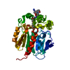 7wamC 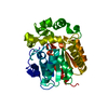 5y2xS S: Starting model for refinement C: citing same article ( |
|---|---|
| Similar structure data |


-Links
 PDBj
PDBj


- Assembly
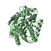
| Deposited unit | 
| ||||||||
|---|---|---|---|---|---|---|---|---|---|
| 1 |
| ||||||||
| Unit cell |
|
-Components
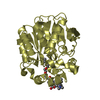
| #1: Protein | Mass: 33716.398 Da / Num. of mol.: 1 Mutation: S2A, L47V, S58T, D78G, Y87F, L88M, C128F, A155T, E160K, A167V, A172T, K175C, C176G, K195N, A224E, N227D, E257K, T264A, H272N, Y273L, P291S, A292T Source method: isolated from a genetically manipulated source Source: (gene. exp.) Rhodococcus sp. (bacteria) / Gene: dhaA / Production host: |
|---|---|
| #2: Chemical | ChemComp-8MS / (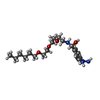  Mass: 384.514 Da / Num. of mol.: 1 / Source method: obtained synthetically / Formula: C19H36N4O4 Mass: 384.514 Da / Num. of mol.: 1 / Source method: obtained synthetically / Formula: C19H36N4O4 |
| #3: Chemical | ChemComp-CL /   Mass: 35.453 Da / Num. of mol.: 1 / Source method: obtained synthetically / Formula: Cl Mass: 35.453 Da / Num. of mol.: 1 / Source method: obtained synthetically / Formula: Cl |
| #4: Water | ChemComp-HOH /  Mass: 18.015 Da / Num. of mol.: 96 / Source method: isolated from a natural source / Formula: H2O Mass: 18.015 Da / Num. of mol.: 96 / Source method: isolated from a natural source / Formula: H2O |
| Has ligand of interest | N |
-Experimental details
-Experiment
| Experiment | Method: X-RAY DIFFRACTION / Number of used crystals: 1 |
|---|
- Sample preparation
| Crystal | Density Matthews: 1.99 Å3/Da / Density % sol: 38.09 % |
|---|---|
| Crystal grow | Temperature: 293 K / Method: vapor diffusion, hanging drop / Details: 25% PEG 8K, 0.1 M MES pH 6.5, 0.2 M Na-acetate |
-Data collection
| Diffraction | Mean temperature: 100 K / Serial crystal experiment: N |
|---|---|
| Diffraction source | Source: SYNCHROTRON / Site: PAL/PLS  / Beamline: 7A (6B, 6C1) / Wavelength: 0.97933 Å / Beamline: 7A (6B, 6C1) / Wavelength: 0.97933 Å |
| Detector | Type: ADSC QUANTUM 270 / Detector: CCD / Date: Apr 2, 2016 |
| Radiation | Protocol: SINGLE WAVELENGTH / Monochromatic (M) / Laue (L): M / Scattering type: x-ray |
| Radiation wavelength | Wavelength: 0.97933 Å / Relative weight: 1 |
| Reflection | Resolution: 2.28→50 Å / Num. obs: 11723 / % possible obs: 97.9 % / Redundancy: 3 % / Rmerge(I) obs: 0.124 / Net I/σ(I): 12.8 |
| Reflection shell | Resolution: 2.3→2.34 Å / Rmerge(I) obs: 0.289 / Num. unique obs: 551 |
- Processing
| Software |
| ||||||||||||||||||||||||||||||
|---|---|---|---|---|---|---|---|---|---|---|---|---|---|---|---|---|---|---|---|---|---|---|---|---|---|---|---|---|---|---|---|
| Refinement | Method to determine structure: MOLECULAR REPLACEMENT Starting model: 5Y2X Resolution: 2.284→32.284 Å / SU ML: 0.23 / Cross valid method: THROUGHOUT / σ(F): 1.4 / Phase error: 22.25 / Stereochemistry target values: ML
| ||||||||||||||||||||||||||||||
| Solvent computation | Shrinkage radii: 0.9 Å / VDW probe radii: 1.11 Å / Solvent model: FLAT BULK SOLVENT MODEL | ||||||||||||||||||||||||||||||
| Displacement parameters | Biso max: 67.98 Å2 / Biso mean: 21.4749 Å2 / Biso min: 10.62 Å2 | ||||||||||||||||||||||||||||||
| Refinement step | Cycle: final / Resolution: 2.284→32.284 Å
| ||||||||||||||||||||||||||||||
| LS refinement shell | Refine-ID: X-RAY DIFFRACTION / Rfactor Rfree error: 0
|
