 Movie
Movie Controller


+ Open data

- Basic information
| Entry | Database: PDB / ID: 5y2x | ||||||
|---|---|---|---|---|---|---|---|
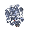
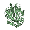
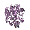
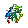
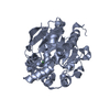
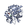
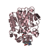
| Title | Crystal structure of apo-HaloTag (M175C) | ||||||
 Components Components | Haloalkane dehalogenase | ||||||
 Keywords Keywords | HYDROLASE / halotag / haloalkane dehalogenase | ||||||
| Function / homology |  Function and homology information Function and homology informationhaloalkane dehalogenase / haloalkane dehalogenase activity / response to toxic substance / membrane Similarity search - Function | ||||||
| Biological species |  Rhodococcus sp. (bacteria) Rhodococcus sp. (bacteria) | ||||||
| Method |  X-RAY DIFFRACTION / SYNCHROTRON / MOLECULAR REPLACEMENT / Resolution: 2.02 Å X-RAY DIFFRACTION / SYNCHROTRON / MOLECULAR REPLACEMENT / Resolution: 2.02 Å | ||||||
 Authors Authors | Lee, H. / Kang, M. / Rhee, H. / Lee, C. | ||||||
 Citation Citation | Journal: Chem. Commun. (Camb.) / Year: 2017 Title: Structure-guided synthesis of a protein-based fluorescent sensor for alkyl halides Authors: Kang, M.G. / Lee, H. / Kim, B.H. / Dunbayev, Y. / Seo, J.K. / Lee, C. / Rhee, H.W. | ||||||
| History |
|
- Structure visualization
| Structure viewer | Molecule: MolmilJmol/JSmol |
|---|
- Downloads & links
-Download
| PDBx/mmCIF format | 5y2x.cif.gz | 79.3 KB | Display | PDBx/mmCIF format |
|---|---|---|---|---|
| PDB format | pdb5y2x.ent.gz | 56.8 KB | Display | PDB format |
| PDBx/mmJSON format | 5y2x.json.gz | Tree view | PDBx/mmJSON format | |
| Others |  Other downloads Other downloads |
-Validation report
| Arichive directory | https://data.pdbj.org/pub/pdb/validation_reports/y2/5y2xftp://data.pdbj.org/pub/pdb/validation_reports/y2/5y2x | HTTPS FTP |
|---|
-Related structure data
| Related structure data | 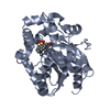 5y2yC 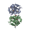 4kafS S: Starting model for refinement C: citing same article ( |
|---|---|
| Similar structure data |


-Links
 PDBj
PDBj


- Assembly
| Deposited unit | 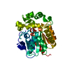
| ||||||||
|---|---|---|---|---|---|---|---|---|---|
| 1 |
| ||||||||
| Unit cell |
|
-Components
| #1: Protein | Mass: 33716.398 Da / Num. of mol.: 1 / Fragment: UNP residues 2-293 Mutation: S2A, L47V, S58T, D78G, Y87F, L88M, C128F, A155T, E160K, A167V, A172T, K175C, C176G, K195N, A224E, N227D, E257K, T264A, H272N, Y273L, P291S, A292T Source method: isolated from a genetically manipulated source Source: (gene. exp.) Rhodococcus sp. (bacteria) / Gene: dhaA / Production host: |
|---|---|
| #2: Chemical | ChemComp-CL /   Mass: 35.453 Da / Num. of mol.: 1 / Source method: obtained synthetically / Formula: Cl Mass: 35.453 Da / Num. of mol.: 1 / Source method: obtained synthetically / Formula: Cl |
| #3: Water | ChemComp-HOH /  Mass: 18.015 Da / Num. of mol.: 259 / Source method: isolated from a natural source / Formula: H2O Mass: 18.015 Da / Num. of mol.: 259 / Source method: isolated from a natural source / Formula: H2O |
-Experimental details
-Experiment
| Experiment | Method: X-RAY DIFFRACTION / Number of used crystals: 1 |
|---|
- Sample preparation
| Crystal | Density Matthews: 2.39 Å3/Da / Density % sol: 48.61 % |
|---|---|
| Crystal grow | Temperature: 293 K / Method: vapor diffusion, hanging drop Details: 25% PEG 20K, 0.1M Tris pH 8.2, 200mM MgCl2, 5% butanol. |
-Data collection
| Diffraction | Mean temperature: 100 K |
|---|---|
| Diffraction source | Source: SYNCHROTRON / Site: PAL/PLS  / Beamline: 7A (6B, 6C1) / Wavelength: 1 Å / Beamline: 7A (6B, 6C1) / Wavelength: 1 Å |
| Detector | Type: ADSC QUANTUM 270 / Detector: CCD / Date: Dec 14, 2015 |
| Radiation | Protocol: SINGLE WAVELENGTH / Monochromatic (M) / Laue (L): M / Scattering type: x-ray |
| Radiation wavelength | Wavelength: 1 Å / Relative weight: 1 |
| Reflection | Resolution: 2.02→50 Å / Num. obs: 22255 / % possible obs: 99.5 % / Redundancy: 6.1 % / Rmerge(I) obs: 0.094 / Net I/σ(I): 26.4 |
| Reflection shell | Rmerge(I) obs: 0.334 |
- Processing
| Software |
| |||||||||||||||||||||
|---|---|---|---|---|---|---|---|---|---|---|---|---|---|---|---|---|---|---|---|---|---|---|
| Refinement | Method to determine structure: MOLECULAR REPLACEMENT Starting model: 4KAF Resolution: 2.02→34.44 Å / Cross valid method: FREE R-VALUE
| |||||||||||||||||||||
| Refinement step | Cycle: LAST / Resolution: 2.02→34.44 Å
|
