[English] 日本語
Yorodumi
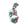
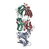
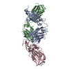
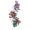
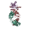
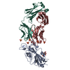
- PDB-7vux: Complex structure of PD1 and 609A-Fab -

+
Open data


ID or keywords:

Loading...

-
Basic information

Entry
Database: PDB / ID: 7vux
TitleComplex structure of PD1 and 609A-Fab
Components
  • Heavy chain of Fab fragment
  • Light chain of Fab fragment
  • Programmed cell death protein 1
KeywordsIMMUNE SYSTEM / PD1 Fab complex
Function / homology
Function and homology information


negative regulation of tolerance induction / regulatory T cell apoptotic process / B cell apoptotic process / negative regulation of immune response / positive regulation of T cell apoptotic process / negative regulation of B cell apoptotic process / humoral immune response / PD-1 signaling / regulation of immune response / adaptive immune response ...negative regulation of tolerance induction / regulatory T cell apoptotic process / B cell apoptotic process / negative regulation of immune response / positive regulation of T cell apoptotic process / negative regulation of B cell apoptotic process / humoral immune response / PD-1 signaling / regulation of immune response / adaptive immune response / Potential therapeutics for SARS / external side of plasma membrane / apoptotic process / plasma membrane
Similarity search - Function
Programmed cell death protein 1 / Immunoglobulin V-Type / Immunoglobulin V-set domain / Immunoglobulin V-set domain / Immunoglobulin subtype / Immunoglobulin / Ig-like domain profile. / Immunoglobulin-like domain / Immunoglobulin-like domain superfamily / Immunoglobulin-like fold
Similarity search - Domain/homology
DI(HYDROXYETHYL)ETHER / THREONINE / Programmed cell death protein 1
Similarity search - Component
Biological speciesHomo sapiens (human)
MethodX-RAY DIFFRACTION / SYNCHROTRON / MOLECULAR REPLACEMENT / molecular replacement / Resolution: 1.64 Å
AuthorsHuang, H. / Zhu, Z. / Zhao, J. / Jiang, L. / Yang, H. / Deng, L. / Meng, X. / Ding, J. / Yang, S. / Zhao, L. ...Huang, H. / Zhu, Z. / Zhao, J. / Jiang, L. / Yang, H. / Deng, L. / Meng, X. / Ding, J. / Yang, S. / Zhao, L. / Xu, W. / Wang, X.
Funding support1items
OrganizationGrant numberCountry
Not funded
CitationJournal: Mabs / Year: 2022
Title: A strategy for the efficient construction of anti-PD1-based bispecific antibodies with desired IgG-like properties.
Authors: Zhao, J. / Jiang, L. / Yang, H. / Deng, L. / Meng, X. / Ding, J. / Yang, S. / Zhao, L. / Xu, W. / Wang, X. / Zhu, Z. / Huang, H.
History
DepositionNov 4, 2021Deposition site: PDBJ / Processing site: PDBJ
Revision 1.0Nov 17, 2021Provider: repository / Type: Initial release
Revision 1.1Mar 16, 2022Group: Database references / Category: citation / citation_author
Item: _citation.country / _citation.journal_abbrev ..._citation.country / _citation.journal_abbrev / _citation.journal_id_CSD / _citation.journal_id_ISSN / _citation.journal_volume / _citation.page_first / _citation.page_last / _citation.pdbx_database_id_DOI / _citation.pdbx_database_id_PubMed / _citation.title / _citation.year / _citation_author.name
Revision 1.2Nov 29, 2023Group: Data collection / Refinement description
Category: chem_comp_atom / chem_comp_bond / pdbx_initial_refinement_model

-
Structure visualization

Structure viewerMolecule:
MolmilJmol/JSmol

Downloads & links

-
Assembly

Deposited unit
A: Programmed cell death protein 1
H: Heavy chain of Fab fragment
L: Light chain of Fab fragment
hetero molecules


Theoretical massNumber of molelcules
Total (without water)63,17822
Polymers61,7903
Non-polymers1,38819
Water14,214789
1


  • Idetical with deposited unit
  • defined by author&software
  • Evidence: gel filtration
TypeNameSymmetry operationNumber
identity operation1_555x,y,z1
Buried area9290 Å2
ΔGint-45 kcal/mol
Surface area25430 Å2
MethodPISA
Unit cell
Length a, b, c (Å)56.290, 68.410, 77.310
Angle α, β, γ (deg.)90.000, 99.440, 90.000
Int Tables number4
Space group name H-MP1211

-
Components

-
Protein , 1 types, 1 molecules A

#1: Protein Programmed cell death protein 1 / Protein PD-1 / hPD-1


Mass: 15313.052 Da / Num. of mol.: 1
Source method: isolated from a genetically manipulated source
Source: (gene. exp.) Homo sapiens (human) / Gene: PDCD1, PD1 / Production host: Escherichia coli (E. coli) / References: UniProt: Q15116

-
Antibody , 2 types, 2 molecules HL

#2: Antibody Heavy chain of Fab fragment


Mass: 23037.746 Da / Num. of mol.: 1
Source method: isolated from a genetically manipulated source
Source: (gene. exp.) Homo sapiens (human)
Production host: mammal environmental sample (environmental samples)
#3: Antibody Light chain of Fab fragment


Mass: 23439.035 Da / Num. of mol.: 1
Source method: isolated from a genetically manipulated source
Source: (gene. exp.) Homo sapiens (human)
Production host: mammal environmental sample (environmental samples)

-
Non-polymers , 7 types, 808 molecules

#4: Chemical ChemComp-THR / THREONINE


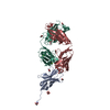
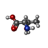
Type: L-peptide linking / Mass: 119.119 Da / Num. of mol.: 1 / Source method: obtained synthetically / Formula: C4H9NO3
#5: Chemical
ChemComp-GOL / GLYCEROL / GLYCERIN / PROPANE-1,2,3-TRIOL


Mass: 92.094 Da / Num. of mol.: 7 / Source method: obtained synthetically / Formula: C3H8O3
#6: Chemical
ChemComp-EDO / 1,2-ETHANEDIOL / ETHYLENE GLYCOL


Mass: 62.068 Da / Num. of mol.: 7 / Source method: obtained synthetically / Formula: C2H6O2
#7: Chemical ChemComp-MG / MAGNESIUM ION


Mass: 24.305 Da / Num. of mol.: 2 / Source method: obtained synthetically / Formula: Mg
#8: Chemical ChemComp-PEG / DI(HYDROXYETHYL)ETHER


Mass: 106.120 Da / Num. of mol.: 1 / Source method: obtained synthetically / Formula: C4H10O3
#9: Chemical ChemComp-CL / CHLORIDE ION


Mass: 35.453 Da / Num. of mol.: 1 / Source method: obtained synthetically / Formula: Cl
#10: Water ChemComp-HOH / water


Mass: 18.015 Da / Num. of mol.: 789 / Source method: isolated from a natural source / Formula: H2O

-
Details

Has ligand of interestN

-
Experimental details

-
Experiment

ExperimentMethod: X-RAY DIFFRACTION / Number of used crystals: 1

-
Sample preparation

CrystalDensity Matthews: 2.44 Å3/Da / Density % sol: 49.56 %
Crystal growTemperature: 291.15 K / Method: vapor diffusion, sitting drop / Details: 0.2 M Ammonium formate, pH 6.6, 20% w/v PEG 3350

-
Data collection

DiffractionMean temperature: 80 K / Serial crystal experiment: N
Diffraction sourceSource: SYNCHROTRON / Site: SSRF / Beamline: BL18U1 / Wavelength: 0.97915 Å
DetectorType: DECTRIS PILATUS3 6M / Detector: PIXEL / Date: May 9, 2020
RadiationProtocol: SINGLE WAVELENGTH / Monochromatic (M) / Laue (L): M / Scattering type: x-ray
Radiation wavelengthWavelength: 0.97915 Å / Relative weight: 1
ReflectionResolution: 1.64→48.91 Å / Num. obs: 70749 / % possible obs: 99.7 % / Redundancy: 3.3 % / CC1/2: 0.996 / Rmerge(I) obs: 0.049 / Rpim(I) all: 0.045 / Rrim(I) all: 0.067 / Net I/σ(I): 12.5
Reflection shellResolution: 1.64→1.67 Å / Redundancy: 3.4 % / Rmerge(I) obs: 0.457 / Mean I/σ(I) obs: 2.2 / Num. unique obs: 3505 / CC1/2: 0.842 / Rpim(I) all: 0.413 / Rrim(I) all: 0.618 / % possible all: 99.9

-
Phasing

PhasingMethod: molecular replacement

-
Processing

Software
NameVersionClassificationNB
XDSdata reduction
Aimlessdata scaling
PHASERphasing
REFMAC5.8.0258refinement
PDB_EXTRACT3.27data extraction
RefinementMethod to determine structure: MOLECULAR REPLACEMENT
Starting model: 5WT9
Resolution: 1.64→48.91 Å / Cor.coef. Fo:Fc: 0.971 / Cor.coef. Fo:Fc free: 0.954 / SU B: 1.959 / SU ML: 0.065 / Cross valid method: THROUGHOUT / σ(F): 0 / ESU R: 0.092 / ESU R Free: 0.096 / Stereochemistry target values: MAXIMUM LIKELIHOOD
Details: HYDROGENS HAVE BEEN ADDED IN THE RIDING POSITIONS U VALUES : REFINED INDIVIDUALLY
RfactorNum. reflection% reflectionSelection details
Rfree0.2019 3529 5 %RANDOM
Rwork0.1596 ---
obs0.1617 67222 99.69 %-
Solvent computationIon probe radii: 0.8 Å / Shrinkage radii: 0.8 Å / VDW probe radii: 1.2 Å / Solvent model: MASK
Displacement parametersBiso max: 104.66 Å2 / Biso mean: 20.227 Å2 / Biso min: 8.98 Å2
Baniso -1Baniso -2Baniso -3
1-0.74 Å20 Å2-0.43 Å2
2---0.07 Å20 Å2
3----0.5 Å2
Refinement stepCycle: final / Resolution: 1.64→48.91 Å
ProteinNucleic acidLigandSolventTotal
Num. atoms4227 0 94 789 5110
Biso mean--36.44 34.03 -
Num. residues----551
Refine LS restraints
Refine-IDTypeDev idealDev ideal targetNumber
X-RAY DIFFRACTIONr_bond_refined_d0.0130.0134751
X-RAY DIFFRACTIONr_bond_other_d0.0010.0174238
X-RAY DIFFRACTIONr_angle_refined_deg1.7841.6476492
X-RAY DIFFRACTIONr_angle_other_deg1.4721.5729907
X-RAY DIFFRACTIONr_dihedral_angle_1_deg20.7135.402647
X-RAY DIFFRACTIONr_dihedral_angle_2_deg31.29221.435216
X-RAY DIFFRACTIONr_dihedral_angle_3_deg12.23515735
X-RAY DIFFRACTIONr_dihedral_angle_4_deg17.8311531
X-RAY DIFFRACTIONr_chiral_restr0.0820.2615
X-RAY DIFFRACTIONr_gen_planes_refined0.010.026281
X-RAY DIFFRACTIONr_gen_planes_other0.0030.021028
LS refinement shellResolution: 1.64→1.683 Å / Rfactor Rfree error: 0 / Total num. of bins used: 20
RfactorNum. reflection% reflection
Rfree0.453 263 -
Rwork0.407 4947 -
all-5210 -
obs--99.87 %

+
About Yorodumi

-
News

-
Feb 9, 2022. New format data for meta-information of EMDB entries

New format data for meta-information of EMDB entries

  • Version 3 of the EMDB header file is now the official format.
  • The previous official version 1.9 will be removed from the archive.

Related info.:EMDB header

External links:wwPDB to switch to version 3 of the EMDB data model

-
Aug 12, 2020. Covid-19 info

Covid-19 info

URL: https://pdbjlvh1.pdbj.org/emnavi/covid19.php

New page: Covid-19 featured information page in EM Navigator.

Related info.:Covid-19 info / Mar 5, 2020. Novel coronavirus structure data

+
Mar 5, 2020. Novel coronavirus structure data

Novel coronavirus structure data

Related info.:Yorodumi Speices / Aug 12, 2020. Covid-19 info

External links:COVID-19 featured content - PDBj / Molecule of the Month (242):Coronavirus Proteases

+
Jan 31, 2019. EMDB accession codes are about to change! (news from PDBe EMDB page)

EMDB accession codes are about to change! (news from PDBe EMDB page)

  • The allocation of 4 digits for EMDB accession codes will soon come to an end. Whilst these codes will remain in use, new EMDB accession codes will include an additional digit and will expand incrementally as the available range of codes is exhausted. The current 4-digit format prefixed with “EMD-” (i.e. EMD-XXXX) will advance to a 5-digit format (i.e. EMD-XXXXX), and so on. It is currently estimated that the 4-digit codes will be depleted around Spring 2019, at which point the 5-digit format will come into force.
  • The EM Navigator/Yorodumi systems omit the EMD- prefix.

Related info.:Q: What is EMD? / ID/Accession-code notation in Yorodumi/EM Navigator

External links:EMDB Accession Codes are Changing Soon! / Contact to PDBj

+
Jul 12, 2017. Major update of PDB

Major update of PDB

  • wwPDB released updated PDB data conforming to the new PDBx/mmCIF dictionary.
  • This is a major update changing the version number from 4 to 5, and with Remediation, in which all the entries are updated.
  • In this update, many items about electron microscopy experimental information are reorganized (e.g. em_software).
  • Now, EM Navigator and Yorodumi are based on the updated data.

External links:wwPDB Remediation / Enriched Model Files Conforming to OneDep Data Standards Now Available in the PDB FTP Archive

-
Yorodumi

Thousand views of thousand structures

  • Yorodumi is a browser for structure data from EMDB, PDB, SASBDB, etc.
  • This page is also the successor to EM Navigator detail page, and also detail information page/front-end page for Omokage search.
  • The word "yorodu" (or yorozu) is an old Japanese word meaning "ten thousand". "mi" (miru) is to see.

Related info.:EMDB / PDB / SASBDB / Comparison of 3 databanks / Yorodumi Search / Aug 31, 2016. New EM Navigator & Yorodumi / Yorodumi Papers / Jmol/JSmol / Function and homology information / Changes in new EM Navigator and Yorodumi

Read more