 Movie
Movie Controller

[English] 日本語
 Yorodumi
- PDB-7vn9: Crystal structure of human coronavirus 229E spike protein recepto... -
+ Open data

- Basic information
| Entry | Database: PDB / ID: 7vn9 | ||||||
|---|---|---|---|---|---|---|---|
| Title | Crystal structure of human coronavirus 229E spike protein receptor-binding domain in complex with C04 Fab | ||||||
 Components Components |
| ||||||
 Keywords Keywords | VIRAL PROTEIN/IMMUNE SYSTEM / Neutralizing antibody / human coronavirus 229E / C04 Fab. / IMMUNE SYSTEM / VIRAL PROTEIN-IMMUNE SYSTEM complex | ||||||
| Function / homology |  Function and homology information Function and homology informationhost cell endoplasmic reticulum-Golgi intermediate compartment membrane / receptor-mediated virion attachment to host cell / endocytosis involved in viral entry into host cell / fusion of virus membrane with host plasma membrane / fusion of virus membrane with host endosome membrane / viral envelope / virion membrane / membrane Similarity search - Function | ||||||
| Biological species |  Homo sapiens (human) Homo sapiens (human) Human coronavirus 229E Human coronavirus 229E | ||||||
| Method |  X-RAY DIFFRACTION / SYNCHROTRON / MOLECULAR REPLACEMENT / Resolution: 4.49 Å X-RAY DIFFRACTION / SYNCHROTRON / MOLECULAR REPLACEMENT / Resolution: 4.49 Å | ||||||
 Authors Authors | Xiang, J.C. / Yang, B. | ||||||
| Funding support |  China, 1items China, 1items
| ||||||
 Citation Citation | Journal: Commun Biol / Year: 2022 Title: Antigenic mapping reveals sites of vulnerability on alpha-HCoV spike protein. Authors: Xiang, J. / Su, J. / Lan, Q. / Zhao, W. / Zhou, Y. / Xu, Y. / Niu, J. / Xia, S. / Qi, Q. / Sidhu, S. / Lu, L. / Miersch, S. / Yang, B. | ||||||
| History |
|
- Structure visualization
| Structure viewer | Molecule: MolmilJmol/JSmol |
|---|
- Downloads & links
-Download
| PDBx/mmCIF format | 7vn9.cif.gz | 442.4 KB | Display | PDBx/mmCIF format |
|---|---|---|---|---|
| PDB format | pdb7vn9.ent.gz | 369.2 KB | Display | PDB format |
| PDBx/mmJSON format | 7vn9.json.gz | Tree view | PDBx/mmJSON format | |
| Others |  Other downloads Other downloads |
-Validation report
| Arichive directory | https://data.pdbj.org/pub/pdb/validation_reports/vn/7vn9ftp://data.pdbj.org/pub/pdb/validation_reports/vn/7vn9 | HTTPS FTP |
|---|
-Related structure data
| Related structure data |  7vmzC 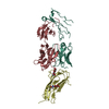 7vngC 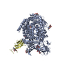 6atkS  6df2S S: Starting model for refinement C: citing same article ( |
|---|---|
| Similar structure data |
-Links
 PDBj
PDBj


- Assembly
| Deposited unit | 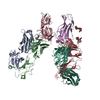
| |||||||||||||||||||||||||||||||||||||||||||||||||||||||||||||||||||||||||
|---|---|---|---|---|---|---|---|---|---|---|---|---|---|---|---|---|---|---|---|---|---|---|---|---|---|---|---|---|---|---|---|---|---|---|---|---|---|---|---|---|---|---|---|---|---|---|---|---|---|---|---|---|---|---|---|---|---|---|---|---|---|---|---|---|---|---|---|---|---|---|---|---|---|---|
| 1 | 
| |||||||||||||||||||||||||||||||||||||||||||||||||||||||||||||||||||||||||
| 2 | 
| |||||||||||||||||||||||||||||||||||||||||||||||||||||||||||||||||||||||||
| Unit cell |
| |||||||||||||||||||||||||||||||||||||||||||||||||||||||||||||||||||||||||
| Noncrystallographic symmetry (NCS) | NCS domain:
NCS domain segments:
|