 Movie
Movie Controller

[English] 日本語
 Yorodumi
- PDB-7vng: Crystal structure of human coronavirus 229E spike protein recepto... -
+ Open data

- Basic information
| Entry | Database: PDB / ID: 7vng | ||||||
|---|---|---|---|---|---|---|---|
| Title | Crystal structure of human coronavirus 229E spike protein receptor-binding domain in complex with S11 Fab | ||||||
 Components Components |
| ||||||
 Keywords Keywords | VIRAL PROTEIN/IMMUNE SYSTEM / human coronavirus 229E / spike protein / RBD / antibody / S11 / IMMUNE SYSTEM / VIRAL PROTEIN-IMMUNE SYSTEM complex | ||||||
| Function / homology |  Function and homology information Function and homology informationhost cell endoplasmic reticulum-Golgi intermediate compartment membrane / receptor-mediated virion attachment to host cell / endocytosis involved in viral entry into host cell / fusion of virus membrane with host plasma membrane / fusion of virus membrane with host endosome membrane / viral envelope / virion membrane / membrane Similarity search - Function | ||||||
| Biological species |  Human coronavirus 229E Human coronavirus 229E Homo sapiens (human) Homo sapiens (human) | ||||||
| Method |  X-RAY DIFFRACTION / SYNCHROTRON / MOLECULAR REPLACEMENT / Resolution: 3.8 Å X-RAY DIFFRACTION / SYNCHROTRON / MOLECULAR REPLACEMENT / Resolution: 3.8 Å | ||||||
 Authors Authors | Xiang, J.C. / Zhao, W.W. / Yang, B. | ||||||
| Funding support |  China, 1items China, 1items
| ||||||
 Citation Citation | Journal: Commun Biol / Year: 2022 Title: Antigenic mapping reveals sites of vulnerability on alpha-HCoV spike protein. Authors: Xiang, J. / Su, J. / Lan, Q. / Zhao, W. / Zhou, Y. / Xu, Y. / Niu, J. / Xia, S. / Qi, Q. / Sidhu, S. / Lu, L. / Miersch, S. / Yang, B. | ||||||
| History |
|
- Structure visualization
| Structure viewer | Molecule: MolmilJmol/JSmol |
|---|
- Downloads & links
-Download
| PDBx/mmCIF format | 7vng.cif.gz | 258.8 KB | Display | PDBx/mmCIF format |
|---|---|---|---|---|
| PDB format | pdb7vng.ent.gz | 176.8 KB | Display | PDB format |
| PDBx/mmJSON format | 7vng.json.gz | Tree view | PDBx/mmJSON format | |
| Others |  Other downloads Other downloads |
-Validation report
| Arichive directory | https://data.pdbj.org/pub/pdb/validation_reports/vn/7vngftp://data.pdbj.org/pub/pdb/validation_reports/vn/7vng | HTTPS FTP |
|---|
-Related structure data
| Related structure data |  7vmzC 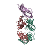 7vn9SC S: Starting model for refinement C: citing same article ( |
|---|---|
| Similar structure data |
-Links
 PDBj
PDBj

- Assembly
| Deposited unit | 
| ||||||||||||
|---|---|---|---|---|---|---|---|---|---|---|---|---|---|
| 1 |
| ||||||||||||
| Unit cell |
|
-Components
| #1: Protein | Mass: 16194.324 Da / Num. of mol.: 1 / Fragment: receptor binding domain Source method: isolated from a genetically manipulated source Source: (gene. exp.) Human coronavirus 229E / Gene: S, 2 / Production host:  Trichoplusia ni (cabbage looper) / References: UniProt: P15423 Trichoplusia ni (cabbage looper) / References: UniProt: P15423 | ||||
|---|---|---|---|---|---|
| #2: Antibody | Mass: 23277.957 Da / Num. of mol.: 1 Source method: isolated from a genetically manipulated source Source: (gene. exp.) Homo sapiens (human)Production host:  | ||||
| #3: Antibody | Mass: 23345.889 Da / Num. of mol.: 1 Source method: isolated from a genetically manipulated source Source: (gene. exp.) Homo sapiens (human)Production host: | ||||
| #4: Sugar |   Type: D-saccharide, beta linking / Mass: 221.208 Da / Num. of mol.: 2 / Source method: obtained synthetically / Formula: C8H15NO6 Type: D-saccharide, beta linking / Mass: 221.208 Da / Num. of mol.: 2 / Source method: obtained synthetically / Formula: C8H15NO6Has ligand of interest | N | Has protein modification | Y | |
-Experimental details
-Experiment
| Experiment | Method: X-RAY DIFFRACTION / Number of used crystals: 1 |
|---|
- Sample preparation
| Crystal | Density Matthews: 3.85 Å3/Da / Density % sol: 68.02 % / Description: block |
|---|---|
| Crystal grow | Temperature: 293 K / Method: vapor diffusion, hanging drop / pH: 7 / Details: 3.6M Sodium Formate, 10% Glycerol |
-Data collection
| Diffraction | Mean temperature: 80 K / Serial crystal experiment: N |
|---|---|
| Diffraction source | Source: SYNCHROTRON / Site: SSRF / Beamline: BL19U1 / Wavelength: 0.978 Å |
| Detector | Type: DECTRIS PILATUS3 6M / Detector: PIXEL / Date: Jan 14, 2021 |
| Radiation | Protocol: SINGLE WAVELENGTH / Monochromatic (M) / Laue (L): M / Scattering type: x-ray |
| Radiation wavelength | Wavelength: 0.978 Å / Relative weight: 1 |
| Reflection | Resolution: 3.65→68.1 Å / Num. obs: 20687 / % possible obs: 100 % / Redundancy: 12.5 % / Biso Wilson estimate: 154.9 Å2 / CC1/2: 0.998 / Rmerge(I) obs: 0.203 / Rpim(I) all: 0.059 / Rrim(I) all: 0.212 / Net I/σ(I): 7.9 |
| Reflection shell | Resolution: 3.65→4 Å / Redundancy: 13.1 % / Rmerge(I) obs: 3.116 / Mean I/σ(I) obs: 0.9 / Num. unique obs: 2600 / CC1/2: 0.546 / Rpim(I) all: 1.229 / Rrim(I) all: 3.242 / % possible all: 100 |
- Processing
| Software |
| |||||||||||||||||||||||||||||||||||||||||||||||||
|---|---|---|---|---|---|---|---|---|---|---|---|---|---|---|---|---|---|---|---|---|---|---|---|---|---|---|---|---|---|---|---|---|---|---|---|---|---|---|---|---|---|---|---|---|---|---|---|---|---|---|
| Refinement | Method to determine structure: MOLECULAR REPLACEMENT Starting model: 7VN9 Resolution: 3.8→49.07 Å / SU ML: 0.6579 / Cross valid method: FREE R-VALUE / σ(F): 1.34 / Phase error: 37.5043 Stereochemistry target values: GeoStd + Monomer Library + CDL v1.2
| |||||||||||||||||||||||||||||||||||||||||||||||||
| Solvent computation | Shrinkage radii: 0.9 Å / VDW probe radii: 1.11 Å / Solvent model: FLAT BULK SOLVENT MODEL | |||||||||||||||||||||||||||||||||||||||||||||||||
| Displacement parameters | Biso mean: 182.38 Å2 | |||||||||||||||||||||||||||||||||||||||||||||||||
| Refinement step | Cycle: LAST / Resolution: 3.8→49.07 Å
| |||||||||||||||||||||||||||||||||||||||||||||||||
| Refine LS restraints |
| |||||||||||||||||||||||||||||||||||||||||||||||||
| LS refinement shell |
| |||||||||||||||||||||||||||||||||||||||||||||||||
| Refinement TLS params. | Method: refined / Origin x: 2.63474504498 Å / Origin y: 60.272840023 Å / Origin z: 23.4260412009 Å
| |||||||||||||||||||||||||||||||||||||||||||||||||
| Refinement TLS group | Selection details: all |
