 Movie
Movie Controller

[English] 日本語
 Yorodumi
- PDB-7vey: Crystal structure of Cyclosorus parasiticus chalcone synthase 1 (... -
+ Open data

- Basic information
| Entry | Database: PDB / ID: 7vey | ||||||||||||
|---|---|---|---|---|---|---|---|---|---|---|---|---|---|
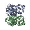
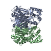
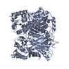
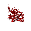
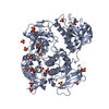
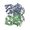
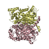
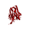
| Title | Crystal structure of Cyclosorus parasiticus chalcone synthase 1 (CpCHS1) | ||||||||||||
 Components Components | chalcone synthases | ||||||||||||
 Keywords Keywords | TRANSFERASE / flavonoids biosynthesis / chalcone synthase / Cyclosorus parasiticus | ||||||||||||
| Function / homology | Thiolase/Chalcone synthase / Peroxisomal Thiolase; Chain A, domain 1 / 3-Layer(aba) Sandwich / Alpha Beta Function and homology information Function and homology information | ||||||||||||
| Biological species |  Cyclosorus parasiticus (plant) Cyclosorus parasiticus (plant) | ||||||||||||
| Method |  X-RAY DIFFRACTION / SYNCHROTRON / MOLECULAR REPLACEMENT / Resolution: 1.9 Å X-RAY DIFFRACTION / SYNCHROTRON / MOLECULAR REPLACEMENT / Resolution: 1.9 Å | ||||||||||||
 Authors Authors | Li, J.X. / Cheng, A.X. | ||||||||||||
| Funding support |  China, 3items China, 3items
| ||||||||||||
 Citation Citation | Journal: Front Plant Sci / Year: 2021 Title: Functional and Structural Investigation of Chalcone Synthases Based on Integrated Metabolomics and Transcriptome Analysis on Flavonoids and Anthocyanins Biosynthesis of the Fern Cyclosorus parasiticus . Authors: Niu, M. / Fu, J. / Ni, R. / Xiong, R.L. / Zhu, T.T. / Lou, H.X. / Zhang, P. / Li, J. / Cheng, A.X. | ||||||||||||
| History |
|
- Structure visualization
| Structure viewer | Molecule: MolmilJmol/JSmol |
|---|
- Downloads & links
-Download
| PDBx/mmCIF format | 7vey.cif.gz | 328 KB | Display | PDBx/mmCIF format |
|---|---|---|---|---|
| PDB format | pdb7vey.ent.gz | 263.6 KB | Display | PDB format |
| PDBx/mmJSON format | 7vey.json.gz | Tree view | PDBx/mmJSON format | |
| Others |  Other downloads Other downloads |
-Validation report
| Arichive directory | https://data.pdbj.org/pub/pdb/validation_reports/ve/7veyftp://data.pdbj.org/pub/pdb/validation_reports/ve/7vey | HTTPS FTP |
|---|
-Related structure data
| Related structure data |  7vezC  7vf0C  6dxbS S: Starting model for refinement C: citing same article ( |
|---|---|
| Similar structure data |

-Links
 PDBj
PDBj- Assembly
| Deposited unit | 
| ||||||||
|---|---|---|---|---|---|---|---|---|---|
| 1 | 
| ||||||||
| 2 | 
| ||||||||
| Unit cell |
|
-Components
| #1: Protein | Mass: 44342.887 Da / Num. of mol.: 4 Source method: isolated from a genetically manipulated source Source: (gene. exp.) Cyclosorus parasiticus (plant) / Production host:  #2: Water | ChemComp-HOH / |  Mass: 18.015 Da / Num. of mol.: 1099 / Source method: isolated from a natural source / Formula: H2O Mass: 18.015 Da / Num. of mol.: 1099 / Source method: isolated from a natural source / Formula: H2O |
|---|
-Experimental details
-Experiment
| Experiment | Method: X-RAY DIFFRACTION / Number of used crystals: 1 |
|---|
- Sample preparation
| Crystal | Density Matthews: 2.41 Å3/Da / Density % sol: 48.91 % |
|---|---|
| Crystal grow | Temperature: 293 K / Method: vapor diffusion, sitting drop / pH: 8 Details: 0.2 M sodium chloride, 0.1 M Tris-HCl, 20 % w/v PEG 4000 |
-Data collection
| Diffraction | Mean temperature: 100 K / Serial crystal experiment: N |
|---|---|
| Diffraction source | Source: SYNCHROTRON / Site: SSRF / Beamline: BL19U1 / Wavelength: 0.9798 Å |
| Detector | Type: DECTRIS PILATUS 6M / Detector: PIXEL / Date: Oct 22, 2020 |
| Radiation | Protocol: SINGLE WAVELENGTH / Monochromatic (M) / Laue (L): M / Scattering type: x-ray |
| Radiation wavelength | Wavelength: 0.9798 Å / Relative weight: 1 |
| Reflection | Resolution: 1.9→41.123 Å / Num. obs: 197428 / % possible obs: 99.4 % / Redundancy: 12.4 % / CC1/2: 0.975 / Net I/σ(I): 10.96 |
| Reflection shell | Resolution: 1.9→1.968 Å / Num. unique obs: 12527 / CC1/2: 0.82 |
- Processing
| Software |
| ||||||||||||||||||||||||||||||||||||||||||||||||||||||||||||||||||||||||||||||||||||||||||||||||||||||||||||||||||||||||||||||||||||
|---|---|---|---|---|---|---|---|---|---|---|---|---|---|---|---|---|---|---|---|---|---|---|---|---|---|---|---|---|---|---|---|---|---|---|---|---|---|---|---|---|---|---|---|---|---|---|---|---|---|---|---|---|---|---|---|---|---|---|---|---|---|---|---|---|---|---|---|---|---|---|---|---|---|---|---|---|---|---|---|---|---|---|---|---|---|---|---|---|---|---|---|---|---|---|---|---|---|---|---|---|---|---|---|---|---|---|---|---|---|---|---|---|---|---|---|---|---|---|---|---|---|---|---|---|---|---|---|---|---|---|---|---|---|
| Refinement | Method to determine structure: MOLECULAR REPLACEMENT Starting model: 6DXB Resolution: 1.9→41.123 Å / SU ML: 0.17 / Cross valid method: THROUGHOUT / σ(F): 1.34 / Phase error: 21.78 / Stereochemistry target values: ML
| ||||||||||||||||||||||||||||||||||||||||||||||||||||||||||||||||||||||||||||||||||||||||||||||||||||||||||||||||||||||||||||||||||||
| Solvent computation | Shrinkage radii: 0.9 Å / VDW probe radii: 1.11 Å / Solvent model: FLAT BULK SOLVENT MODEL | ||||||||||||||||||||||||||||||||||||||||||||||||||||||||||||||||||||||||||||||||||||||||||||||||||||||||||||||||||||||||||||||||||||
| Displacement parameters | Biso max: 55.28 Å2 / Biso mean: 8.9724 Å2 / Biso min: 1.44 Å2 | ||||||||||||||||||||||||||||||||||||||||||||||||||||||||||||||||||||||||||||||||||||||||||||||||||||||||||||||||||||||||||||||||||||
| Refinement step | Cycle: final / Resolution: 1.9→41.123 Å
| ||||||||||||||||||||||||||||||||||||||||||||||||||||||||||||||||||||||||||||||||||||||||||||||||||||||||||||||||||||||||||||||||||||
| Refine LS restraints |
| ||||||||||||||||||||||||||||||||||||||||||||||||||||||||||||||||||||||||||||||||||||||||||||||||||||||||||||||||||||||||||||||||||||
| LS refinement shell | Refine-ID: X-RAY DIFFRACTION / Rfactor Rfree error: 0
|
